

Efficacy and Safety of a Polytetrafluoroethylene Membrane Wrapped a Single Layer of Sirolimus-Eluting Stent in a Porcine Coronary Perforation Model
- PMID: 39076929
- PMCID: PMC11266774
- DOI: 10.31083/j.rcm2307233
Efficacy and Safety of a Polytetrafluoroethylene Membrane Wrapped a Single Layer of Sirolimus-Eluting Stent in a Porcine Coronary Perforation Model
Abstract
Background: Covered stents are effective in treating coronary artery perforation (CAP), however, the high rate of immediate device deployment failure and in-stent restenosis have limited the application of the currently covered stents.
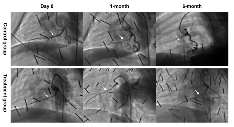
Methods: We designed a covered stent system consisting of a single layer of drug-eluting stent and a layer of polytetrafluoroethylene (PTFE) membrane wrapped at the outer layer of the stent. The immediate sealing effect of our novel covered stent was observed by using an Ellis type III CAP model. The device's success was defined as its ability to seal the perforation, assessed by visual estimation and final thrombolysis in myocardial infarction (TIMI) 3 flow. The antiproliferative effect was evaluated in 12 swine, which were randomly assigned to treatment (sirolimus-eluting covered stents) and control (bare metal covered stents) groups. Coronary angiography and optical coherence tomography (OCT) were performed at index procedure, 1- and 6-month after stent implantation. All swine were sacrificed for histopathological analyses at 6-month.
Results: The device success rate was 100%. All swine were alive at 6-month follow-up. At 1-month, the treatment group had a larger minimal luminal diameter (MLD) (1.89 0.29 mm vs. 0.63 0.65 mm, p = 0.004) and lower late luminal loss (LLL) (0.47 0.15 mm vs. 1.80 0.34 mm, p 0.001) compared with control group. At 6-month, the treatment group had a numerically higher MLD (0.94 0.75 mm vs. 0.26 0.46 mm; p = 0.230) and lower LLL (1.43 0.85 mm vs. 2.17 0.28 mm; p = 0.215) compared with control group. Histological analyses revealed the mean plaque area was lower in the treatment group (2.99 0.81 vs. 4.29 0.77 , p = 0.035) than in the control group. No in-stent thrombosis was observed in either group.
Conclusions: In the porcine model of coronary perforation, the PTFE membrane wrapped sirolimus-eluting stent showed a high device success rate in sealing the perforation. The drug-eluting covered stent demonstrated a relatively sustained antiproliferative effect up to 6 months post-implantation.
Keywords: coronary artery perforation; covered stent; percutaneous coronary intervention.
Copyright: © 2022 The Author(s). Published by IMR Press.
Conflict of interest statement
Yinyi (Liaoning) Biotech Co., Ltd (Dalian, China) kindly assisted us on the design and production of the novel covered stent. We declare that Yinyi (Liaoning) Biotech Co., Ltd had no role in the design of the study; in the collection, analyses, or interpretation of data; in the writing of the manuscript, and in the decision to publish the results. The authors declare no conflicts of interest.
Figures


References
-
- Harnek J, James S, Lagerqvist B. Coronary Artery Perforation and Tamponade—Incidence, Risk Factors, Predictors and Outcomes from 12 Years’ Data of the SCAAR Registry. Circulation Journal . 2019;84:43–53. - PubMed
-
- Parikh P, Banerjee K, Sammour Y, Ali AF, Sankaramangalam K, Nair R, et al. Utilization and outcomes of polytetrafluoroethylene covered stents in patients with coronary artery perforation and coronary artery aneurysm: Single center 15-year experience. Catheterization and cardiovascular interventions . 2019;94:555–561. - PubMed
-
- Pavani M, Cerrato E, Latib A, Ryan N, Calcagno S, Rolfo C, et al. Acute and long-term outcomes after polytetrafluoroethylene or pericardium covered stenting for grade 3 coronary artery perforations: Insights from G3-CAP registry. Catheterization and Cardiovascular Interventions . 2018;92:1247–1255. - PubMed
-
- Dogu Kilic I, Fabris E, Serdoz R, Caiazzo G, Foin N, Abou-Sherif S, et al. Coronary covered stents. EuroIntervention . 2016;12:1288–1295. - PubMed
-
- Al-Lamee R, Ielasi A, Latib A, Godino C, Ferraro M, Mussardo M, et al. Incidence, Predictors, Management, Immediate and Long-Term Outcomes Following Grade III Coronary Perforation. JACC: Cardiovascular Interventions . 2011;4:87–95. - PubMed
LinkOut - more resources
Full Text Sources
Miscellaneous
