. 2024 Jul 15:15:1412693.
doi: 10.3389/fimmu.2024.1412693. eCollection 2024.

Neoadjuvant immunochemotherapy improves clinical outcomes of patients with esophageal cancer by mediating anti-tumor immunity of CD8+ T (Tc1) and CD16+ NK cells

Affiliations

Neoadjuvant immunochemotherapy improves clinical outcomes of patients with esophageal cancer by mediating anti-tumor immunity of CD8+ T (Tc1) and CD16+ NK cells

Yunlong He et al. Front Immunol. .

Abstract

Background: Esophageal cancer (ESCA) is one of the most common tumors in the world, and treatment using neoadjuvant therapy (NT) based on radiotherapy and/or chemotherapy has still unsatisfactory results. Neoadjuvant immunochemotherapy (NICT) has also become an effective treatment strategy nowadays. However, its impact on the tumor microenvironment (TME) and regulatory mechanisms on T cells and NK cells needs to be further elucidated.

Methods: A total of 279 cases of ESCA who underwent surgery alone [non-neoadjuvant therapy (NONE)], neoadjuvant chemotherapy (NCT), and NICT were collected, and their therapeutic effect and survival period were compared. Further, RNA sequencing combined with biological information was used to analyze the expression of immune-related genes. Immunohistochemistry, immunofluorescence, and quantitative real-time PCR (qRT-PCR) were used to verify the activation and infiltration status of CD8+ T and CD16+ NK cells, as well as the function and regulatory pathway of killing tumor cells.

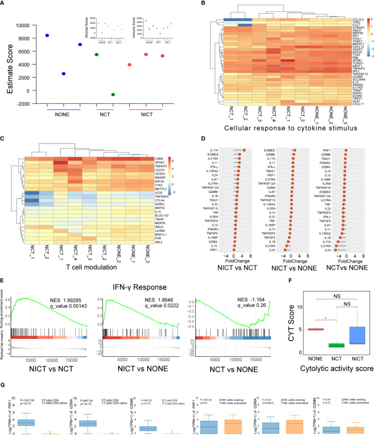
Results: Patients with ESCA in the NICT group showed better clinical response, median survival, and 2-year survival rates (p < 0.05) compared with the NCT group. Our RNA sequencing data revealed that NICT could promote the expression of immune-related genes. The infiltration and activation of immune cells centered with CD8+ T cells were significantly enhanced. CD8+ T cells activated by PD-1 inhibitors secreted more IFN-γ and cytotoxic effector factor cells through the transcription factor of EOMES and TBX21. At the same time, activated CD8+ T cells mediated the CD16+ NK cell activation and secreted more IFN-γ to kill ESCA cells. In addition, the immunofluorescence co-staining results showed that more CD276+ tumor cells and CD16+ NK cells were existed in pre-NCT and pre-NICT group. However, CD276+ tumor cells were reduced significantly in the post-NICT group, while they still appeared in the post-NCT group, which means that CD16+ NK cells can recognize and kill CD276+ tumor cells after immune checkpoint blocker (ICB) treatment.

Conclusion: NICT can improve the therapeutic effect and survival period of resectable ESCA patients. NICT could promote the expression of immune-related genes and activate CD8+ T and CD16+ NK cells to secrete more IFN-γ to kill ESCA cells. It provides a theoretical basis and clinical evidence for its potential as an NT strategy in ESCA.

Keywords: CD16+ NK cells; CD276; CD8+ T cells; esophageal cancer; neoadjuvant immunochemotherapy; tumor microenvironment.


Conflict of interest statement

The authors declare that the research was conducted in the absence of any commercial or financial relationships that could be construed as a potential conflict of interest.

Figures

Figure 1
Figure 1
NICT significantly improved the therapeutic effect of esophageal cancer (ESCA). (A) Survival period. NICT, neoadjuvant immunochemotherapy; NCT, neoadjuvant chemotherapy; HR, hazard ratio. (B) Changes in tumor size. Pre-NCT, pre-neoadjuvant chemotherapy; Post-NCT, post-neoadjuvant chemotherapy; Pre-NICT, pre-neoadjuvant immunochemotherapy; Post-NICT, post-neoadjuvant immunochemotherapy. (C) Conversion rate of clinical stage. (D) The reduction rate of clinical stage. (E) pCR, pathologic complete response. (F) Clinical cases. The maximum longitudinal diameters of tumors in the sagittal plane of CT images. NCT group (left): Case 1 and Case 2 had partial response (PR), and Case 3 had progressive disease (PD). NICT group (right): Case 4 had pCR, and Case 5 and Case 6 had PR. *p < 0.05, **p < 0.01, ***p < 0.001. NS, no significance.
Figure 2
Figure 2
NICT promoted the expression of immune-related genes in ESCA tissues. (A) Pathway enrichment analysis of the cytokine, chemokine, and their receptors in NICT group compared to NCT group using RNA sequencing technique. (B) The function cluster analysis of Gene Ontology (GO) showing comparison of the differential genes between the NCT and NICT groups. (C) The function cluster analysis of GO showing comparison of function clusters of the differential genes between the NONE and NICT groups. (D) Comparison of cycle net plot between the NCT and NICT groups. (E) Comparison of cycle net plot between the NONE and NICT groups. (F) The table presents the tumor sizes pre- and post-treatment for eight clinical cases (from C1 to C8) from the NONE, NCT, and NICT groups. (G) qRT-PCR results of 12 immune-related genes and immune-regulated genes in clinical cases among the NONE, NCT, and NICT groups. NICT, neoadjuvant immunochemotherapy; ESCA, esophageal cancer; NCT, neoadjuvant chemotherapy; NONE, non-neoadjuvant therapy; qRT-PCR, quantitative real-time PCR.
Figure 3
Figure 3
NICT activated the immune microenvironment of ESCA tissues. (A) Using ESTIMATE Score to analyze the scoring of immune cell infiltration among NONE, NCT, and NICT groups. (B) Heatmap showing the production of cellular response to cytokine stimulus in each group of clinical cases. (C) Heatmap showing the production of T-cell modulation in each group of clinical cases. (D) Comparison of the expression of cytokines in the NONE, NCT, and NICT groups, especially the cytotoxic effector factors specifically expressed in T cells and NK cells. (E) Comparison of IFN-γ response pathways among NONE, NCT, and NICT groups by Gene Set Enrichment Analysis (GSEA). (F) Evaluation of the cytotoxic activity of immune cells in tumor tissues by calculating cytolytic activity score (CYT) using PRF1 and GZMM. (G) Using HAP database to identify the types of T cells and NK cells expressing PRF1, GZMA, and GZMM in ESCA. NICT, neoadjuvant immunochemotherapy; ESCA, esophageal cancer; NONE, non-neoadjuvant therapy; NCT, neoadjuvant chemotherapy. *p < 0.05, NS, no significance.
Figure 4
Figure 4
Anti-tumor immunity of CD8+ T cells was boosted by NICT. (A) Immunofluorescence showing the expression status and quantity changes in CD8+ T cells in the NICT group before and after PD-1 inhibitor treatment and cell count analysis. (B) Immunofluorescence showing the expression status and quantity changes in CD8+ T cells in the NCT group before and after chemotherapy and cell count analysis. (C) qRT-PCR showing the expression level of TCIRG1 in each group of clinical cases. (D) HAP database showing the types of T cells expressing TCIRG1 in ESCA. (E) Multiplex immunofluorescence showing the expression status and quantity changes in CD8+ T cells and TCIRG1 among the NICT and NCT groups pre- and post-NT. (F) Multiplex immunofluorescence showing the expression status and quantity changes in CD8+ T cells and IFN-γ among the NICT and NCT groups pre- and post-NT. (G) qRT-PCR showing the expression levels of TBX21, EOMES, IFN-γ, and GZMM in clinical cases among the NONE, NCT, and NICT groups. **p < 0.01. NICT, neoadjuvant immunochemotherapy; qRT-PCR, quantitative real-time PCR; ESCA, esophageal cancer; NCT, neoadjuvant chemotherapy; NT, neoadjuvant therapy; NONE, non-neoadjuvant therapy.
Figure 5
Figure 5
CD16+ NK cells killing tumor cells were promoted depending on NICT. (A) Immunofluorescence showing the expression status and quantity changes in CD16+ NK cells among the NICT and NCT groups pre- and post-NT and cell count analysis. (B) Multiplex immunofluorescence showing the expression status and quantity changes in CD16+ NK cells and IFN-γ among the NICT and NCT groups pre- and post-NT. (C) Multiplex immunofluorescence showing the expression status and quantity changes in CD16+ NK and CD276+ cells among the NICT and NCT groups pre-NT. (D) Multiplex immunofluorescence showing the expression status and quantity changes in CD16+ NK and CD276+ cells among the NICT and NCT groups post-NT. (E) Pattern diagram of NICT activated the immune cells to kill ESCA cells. **p < 0.01. NICT, neoadjuvant immunochemotherapy; NCT, neoadjuvant chemotherapy; NT, neoadjuvant therapy; ESCA, esophageal cancer.

References

    1. Morgan E, Soerjomataram I, Rumgay H, Coleman HG, Thrift AP, Vignat J, et al. . The global landscape of esophageal squamous cell carcinoma and esophageal adenocarcinoma incidence and mortality in 2020 and projections to 2040: new estimates from globocan 2020. Gastroenterology. (2022) 163:649–58 e2. doi: 10.1053/j.gastro.2022.05.054 - DOI - PubMed
    1. Matsuda S, Kitagawa Y, Takemura R, Okui J, Okamura A, Kawakubo H, et al. . Real-world evaluation of the efficacy of neoadjuvant dcf over cf in esophageal squamous cell carcinoma: propensity score-matched analysis from 85 authorized institutes for esophageal cancer in Japan. Ann Surg. (2023) 278:e35–42. doi: 10.1097/SLA.0000000000005533 - DOI - PubMed
    1. Leng XF, Daiko H, Han YT, Mao YS. Optimal preoperative neoadjuvant therapy for resectable locally advanced esophageal squamous cell carcinoma. Ann N Y Acad Sci. (2020) 1482:213–24. doi: 10.1111/nyas.14508 - DOI - PubMed
    1. Duma N, Santana-Davila R, Molina JR. Non-small cell lung cancer: epidemiology, screening, diagnosis, and treatment. Mayo Clin Proc. (2019) 94:1623–40. doi: 10.1016/j.mayocp.2019.01.013 - DOI - PubMed
    1. Li K, Zhang A, Li X, Zhang H, Zhao L. Advances in clinical immunotherapy for gastric cancer. Biochim Biophys Acta Rev Cancer. (2021) 1876:188615. doi: 10.1016/j.bbcan.2021.188615 - DOI - PubMed

MeSH terms

LinkOut - more resources