Review
. 2022 Oct 10;23(10):335.
doi: 10.31083/j.rcm2310335. eCollection 2022 Oct.

New Diagnostic Approach to Arrhythmogenic Cardiomyopathy: The Padua Criteria

Affiliations
Review

New Diagnostic Approach to Arrhythmogenic Cardiomyopathy: The Padua Criteria

Francesca Graziano et al. Rev Cardiovasc Med. .

Abstract

Arrhythmogenic cardiomyopathy (ACM) is a rare heart muscle disease characterized by a progressive fibro-fatty myocardial replacement, ventricular arrhythmias, and increased risk of sudden cardiac death. The first diagnostic criteria were proposed by an International Task Force of experts in 1994 and revised in 2010. At that time, ACM was mainly considered a right ventricle disease, with left ventricle involvement only in the late stages. Since 2010, several pathological and clinical studies using cardiac magnetic resonance (CMR) imaging have allowed to understand the phenotypic expression of the disease and to reach the current idea that ACM may affect both ventricles. Indeed, left ventricular involvement may parallel or exceed right ventricular involvement. The main limitations of the 2010 criteria included the poor sensitivity for left ventricular involvement and the lack of inclusion of tissue characterization by CMR. The 2020 International criteria (the Padua criteria) were developed to overcome these shortcomings. The most important innovations are the introduction of a set of criteria for identifying left ventricular variants and the use of CMR for tissue characterization. Moreover, criteria for right ventricular involvement were also updated taking into account new evidence. According to the number of criteria for right and/or left ventricular involvement, the 2020 Padua criteria allows diagnosing three ACM phenotypic variants: right-dominant, biventricular and left-dominant. This review discusses the evolving approach to diagnosis of ACM, from the 1994 International Criteria to the 2020 Padua criteria.

Keywords: arrhythmogenic cardiomyopathy; cardiac magnetic resonance; cardiomyopathy; diagnosis; ventricular arrhythmias.


Conflict of interest statement

The authors declare no conflict of interest. Alessandro Zorzi is serving as one of the Editorial Board members of this journal. We declare that Alessandro Zorzi had no involvement in the peer review of this article and has no access to information regarding its peer review. Full responsibility for the editorial process for this article was delegated to Stefan Peters.

Figures

Fig. 1.
Fig. 1.
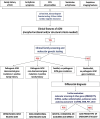
Flowchart for phenotypic characterization of ACM. The diagnosis of ACM requires at least 1 morpho-functional or structural abnormalities criterion, either major or minor. The diagnosis of the specific phenotypic variant depends on the ventricle interested on alterations (see text for details). Moreover, the likelihood of disease is defined by the combination of the major and minor criteria fulfilled. ACM, arrhythmogenic cardiomyopathy; ALVC, arrhythmogenic left ventricular cardiomyopathy; ARVC, arrhythmogenic right ventricular cardiomyopathy; LV, left ventricle; RV, right ventricle. Adapted from Corrado et al. [20].
Fig. 2.
Fig. 2.
Flow-chart for etiology assessment of ACM. After the diagnosis is reached in a proband, cascade family screening and molecular genetic testing may allow to identify patients with identified gene mutation in a desmosomal or non-desmosomal gene. In patients with negative genetic testing, cascade family screening may allow to identify other affected family members: in this case, the diagnosis is a familial disease with still unknown genetic basis (so-called “gene elusive”). In case both genetic testing and family screening are negative, further testing may be performed to exclude phenocopies such as congenital heart disease or myocarditis. Adapted from Corrado et al. [19].
Fig. 3.
Fig. 3.
Clinical features of ARVC. Basal ECG, exercise testing ECG and CMR findings in a 38-year-old woman hospitalized for sustained VT. Basal ECG showed TWI in V1–V5 and flattened T wave in inferior leads (A). Exercise testing revealed frequent PVBs and a non-sustained VT with LBBB/superior axis morphology, originating from RV free wall (B). CMR revealed mild RV dilatation, moderate RV systolic disfunction, a wide peritricuspid aneurysm, with an extreme thinning of the wall (four-chamber cine view in diastolic phase, C, and systolic phase, D). The PD-TSE sequences revealed fatty infiltration of the RV wall, especially in the subtricuspid region (E, and magnified on the top of F). No RV LGE was identified, not even in the same regions of RV fatty infiltration, maybe because an extreme thinning of the RV wall (F on the bottom). The diagnosis was “definite ARVC”. ARVC, arrhythmogenic right ventricular cardiomyopathy; CMR, cardiac magnetic resonance; LBBB, left bundle branch block; LGE, late gadolinium enhancement; PD-TSE, positron density-turbo spin echo; PVBs, premature ventricular beats; RV, right ventricle; TWI, T wave inversion; VT, ventricular tachycardia. Adapted from Graziano et al. [31].
Fig. 4.
Fig. 4.
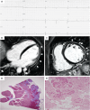
Clinical and histopathological features of ALVC. Basal ECG and CMR findings in a patient who underwent cardiac transplantation because of ALVC related to a DSP gene mutation. Basal ECG revealed low QRS voltages in limb leads and flattened T-waves in infero-lateral leads (A). Post-contrast sequences on CMR (four-chamber view, B, and short-axis view, C) revealed subepicardial LGE involving the anterior septum and the whole LV free wall (“ring like” pattern) from basal to apical regions. Histology in LV inferolateral region demonstrated fibrofatty myocardial replacement in the subepicardial layer (D); a magnification of residual myocytes embedded within fibrous and fatty tissue (hematoxylin and eosin stain) (E). The diagnosis was “definite ALVC”. ALVC, arrhythmogenic left ventricular cardiomyopathy; CMR, cardiac magnetic resonance; DSP, desmoplakin gene; LGE, late gadolinium enhancement; LV, left ventricle. Adapted from Cipriani et al. [14].
Fig. 5.
Fig. 5.
Clinical features of biventricular ACM. Basal ECG and CMR findings in a 28-year-old elite athlete. ECG revealed low QRS voltages in limb leads (A). Exercise testing demonstrated PVBs with a RBBB/superior axis morphology, isolated and in couples. CMR cine-sequences showed hypokinesis of the mid-apical lateral wall and multiple small bulging of the RV free wall (B, four-chamber view). The PD-TSE sequences revealed epicardial fatty infiltration of the lateral and inferior LV walls (C, four-chamber view). Post-contrast sequences on CMR demonstrated a subepicardial stria of LGE involving the antero-lateral, infero-lateral and inferior LV walls (D, short- axis view) and also the RV inferior wall (E, RV inflow-outflow view). The diagnosis was “definite biventricular ACM”. ACM, arrhythmogenic cardiomyopathy; CMR, cardiac magnetic resonance; LV, left ventricle; PVBs, premature ventricular beats; PD-TSE, proton density-turbo spin echo; RBBB, right bundle branch block; RV, right ventricle. Adapted from Zorzi et al. [38].

References

    1. Corrado D, Link MS, Calkins H. Arrhythmogenic Right Ventricular Cardiomyopathy. New England Journal of Medicine . 2017;376:61–72. - PubMed
    1. Maron BJ. Right Ventricular Cardiomyopathy: another Cause of Sudden Death in the Young. New England Journal of Medicine . 1988;318:178–180. - PubMed
    1. Lancisi GM, Caput V, Musca . De motu cordis et aneurysmatibus. Opus posthumu, in duas partes divisum . Naples, Italy: 1736.
    1. Marcus FI, Fontaine GH, Guiraudon G, Frank R, Laurenceau JL, Malergue C, et al. Right ventricular dysplasia: a report of 24 adult cases. Circulation . 1982;65:384–398. - PubMed
    1. Thiene G, Nava A, Corrado D, Rossi L, Pennelli N. Right Ventricular Cardiomyopathy and Sudden Death in Young People. New England Journal of Medicine . 1988;318:129–133. - PubMed

LinkOut - more resources