Review
. 2024 Jan 29;25(2):37.
doi: 10.31083/j.rcm2502037. eCollection 2024 Feb.

Cardiac Sarcoidosis: A Comprehensive Clinical Review

Affiliations
Review

Cardiac Sarcoidosis: A Comprehensive Clinical Review

András Vereckei et al. Rev Cardiovasc Med. .

Abstract

Sarcoidosis is an inflammatory multisystemic disease of unknown etiology characterized by the formation of non-caseating granulomas. Sarcoidosis can affect any organ, predominantly the lungs, lymphatic system, skin and eyes. While > 90% of patients with sarcoidosis have lung involvement, an estimated 5% of patients with sarcoidosis have clinically manifest cardiac sarcoidosis (CS), whereas approximately 25% have asymptomatic, clinically silent cardiac involvement verified by autopsy or imaging studies. CS can present with conduction disturbances, ventricular arrhythmias, heart failure or sudden cardiac death. Approximately 30% of < 60-year-old patients presenting with unexplained high degree atrioventricular (AV) block or ventricular tachycardia are diagnosed with CS, therefore CS should be strongly considered in such patients. CS is the second leading cause of death among patients affected by sarcoidosis after pulmonary sarcoidosis, therefore its early recognition is important, because early treatment may prevent death from cardiovascular involvement. The establishment of isolated CS diagnosis sometimes can be quite difficult, when extracardiac disease cannot be verified. The other reason for the difficulty to diagnose CS is that CS is a chameleon of cardiology and it can mimic (completely or almost completely) different cardiac diseases, such as arrhythmogenic cardiomyopathy, giant cell myocarditis, dilated, restrictive and hypertrophic cardiomyopathies. In this review article we will discuss the current diagnosis and management of CS and delineate the potential difficulties and pitfalls of establishing the diagnosis in atypical cases of isolated CS.

Keywords: cardiac sarcoidosis; granulomatous disease; sarcoidosis.


Conflict of interest statement

The authors declare no conflict of interest.

Figures

Fig. 1.
Fig. 1.
The algorithm devised to distinguish sarcoidosis with left and right ventricular involvement from ACM and its application to the ECG of our patient who had CS mimicking ACM. (A) The ECG algorithm. (B) The application of the algorithm on our patient’s ECG. The PR interval is 220–230 ms, thus already the first step of the algorithm suggests CS. The surface area of the maximum R’ wave in lead V2 marked by light blue color was 1.65 mm2, therefore the third step of the algorithm also suggests CS. R’ wave was defined as any positive deflection after an S wave. Reproduced with permission from [6]. ARVC, arrhythmogenic right ventricular cardiomyopathy; CS, cardiac sarcoidosis; ECG, electrocardiogram; ACM, arrhythmogenic cardiomyopathy.
Fig. 2.
Fig. 2.
CMR images of a 70-year-old female patient with CS. The delayed contrast enhancement images in short axis planes show biventricular late gadolinium enhancement (LGE) corresponding to fibrotic involvement (white arrows) and right ventricular thrombus formation (black arrows). Subepicardial LGE is present in the anterior septum, LV inferior wall, subepicardial-midmyocardial LGE is seen in the LV anterior wall and LGE is present in the RV myocardium in the vicinity of thrombus. CMR, cardiac magnetic resonance; CS, cardiac sarcoidosis; LV, left ventricle; RV, right ventricle.
Fig. 3.
Fig. 3.
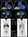
FDG-PET/CT examination of a 65-year-old female patient with histologically (EMB) proven sarcoidosis. Axial fused (A,B,C) and coronal fused (E) PET/CT images and maximum intensity projection (MIP) PET image (D) show increased multifocal FDG uptake in the left ventricular myocardium (A,E) as cardiac involvement, high focal lymph node uptake ((B,D) marked by blue arrows) and pulmonary uptake ((C,D) marked by yellow arrows) indicative for extracardiac manifestation of sarcoidosis. FDG-PET/CT, 18F-fluorodeoxyglucose positron emission tomography/computed tomography; EMB, endomyocardial biopsy.
Fig. 4.
Fig. 4.
Microscopic appearance of sarcoidosis in the endomyocardial biopsy specimen. Top: The non-necrotizing granulomatous inflammation of the myocardium is sharply demarcated, and it is typically surrounded by diffuse fibrosis. The granulomas (- - -) are scattered and typically well circumscribed in sarcoidosis. Diffuse necrosis of cardiomyocytes is absent (H&E staining, 10(×) objective magnification). Bottom: The cellular components of sarcoid granulomas include multinucleated giant cells (Δ), epithelioid macrophages (black arrows) and lymphocytes (white arrows) (H&E staining, 20(×) objective magnification).
Fig. 5.
Fig. 5.
Pre- and posttreatment FDG-PET/CT examination of a 44-year-old male patient with histologically (EMB) proven sarcoidosis. Coronal fused pretreatment and posttreatment (A) and axial fused (B) PET/CT images with volume of interest (VOI) and maximum intensity projection (MIP) PET images before (C) and after immunosuppressive therapy (D) with quantitative parameters. Pretreatment scans show increased multifocal FDG uptake in the left and right ventricular myocardium as cardiac involvement, the presence of high focal supra- and infradiaphragmatic lymph node uptake is indicative of extracardiac sarcoidosis. Posttreatment scans do not show pathological FDG uptake confirming complete treatment response. EMB, endomyocardial biopsy; BW, body weight; FDG-PET/CT, 18F-fluorodeoxyglucose positron emission tomography/computed tomography; PT, positron emission tomography; SUV, standardized uptake values; TLG, total glycolytic activity.
Fig. 6.
Fig. 6.
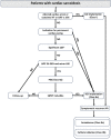
Algorithm for sudden cardiac death prevention and treatment of ventricular arrhythmia in patients with cardiac sarcoidosis. ICD, implantable cardioverter defibrillator; LGE, late gadolinium enhancement; LVEF, left ventricular ejection fraction; PES, programmed electrical stimulation; SMVT, sustained monomorphic ventricular tachycardia; VA, ventricular arrhythmia; VT, ventricular tachycardia. *LGE affecting 9/22 segments or 22% of the LV mass has been associated with arrhythmic endpoints. Reproduced from [79] (Fig. 24 of [79]).

References

    1. Alba AC, Gupta S, Kugathasan L, Ha A, Ochoa A, Balter M, et al. Cardiac Sarcoidosis: A Clinical Overview. Current Problems in Cardiology . 2021;46:100936. - PubMed
    1. Iwai K, Tachibana T, Takemura T, Matsui Y, Kitaichi M, Kawabata Y. Pathological studies on sarcoidosis autopsy. I. Epidemiological features of 320 cases in Japan. Acta Pathologica Japonica . 1993;43:372–376. - PubMed
    1. Perry A, Vuitch F. Causes of death in patients with sarcoidosis. A morphologic study of 38 autopsies with clinicopathologic correlations. Archives of Pathology & Laboratory Medicine . 1995;119:167–172. - PubMed
    1. Drent M, Crouser ED, Grunewald J. Challenges of Sarcoidosis and Its Management. The New England Journal of Medicine . 2021;385:1018–1032. - PubMed
    1. Birnie DH, Kandolin R, Nery PB, Kupari M. Cardiac manifestations of sarcoidosis: diagnosis and management. European Heart Journal . 2017;38:2663–2670. - PubMed

LinkOut - more resources