. 2024 Jul 23:17:4957-4973.
doi: 10.2147/JIR.S464655. eCollection 2024.

Circ_0008146 Exacerbates Ferroptosis via Regulating the miR-342-5p/ACSL4 Axis After Cerebral Ischemic/Reperfusion

Affiliations

Circ_0008146 Exacerbates Ferroptosis via Regulating the miR-342-5p/ACSL4 Axis After Cerebral Ischemic/Reperfusion

Cai-Dong Liu et al. J Inflamm Res. .

Abstract

Purpose: Acute ischemic stroke (AIS) has seriously threatened people's health worldwide and there is an urge need for early diagnosis and effective treatment of AIS. This research intended to clarify the regulatory role of circ_0008146/miR-342-5p/ACSL4 axis in AIS.

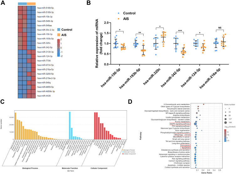
Methods: High-throughput small RNA sequencing analysis was adapted to identify differentially expressed miRNAs between the AIS and control group. The circ_0008146, miR-342-5p, and ACSL4 levels were detected by qRT-PCR. Middle cerebral artery occlusion/reperfusion (MCAO/R) models were constructed in C57BL/6J mice. Assay kits were used to determine Fe2+ levels and a battery of oxidative stress and lipid peroxidation indicators, including ROS, MDA, LPO, SOD and GSH/GSSG ratio. The protein levels of ACSL4 were measured by Western blot. The behavioral function was assessed using neurobehavioral tests. TTC staining was employed to visualize infarction size. Nissl staining was adapted to detect histopathological changes. Receiver operating characteristic curve and correlation analysis were applied to investigate the clinical value and association of miR-342-5p and ACSL4.

Results: A total of 44 AIS patients and 49 healthy controls were enrolled in our study. The small RNA sequencing unveiled a significant decrease in miR-342-5p levels in AIS patients. MiR-342-5p inhibited oxidative stress and RSL3-induced ferroptosis after cerebral ischemic/reperfusion injury in vivo by targeting ferroptosis-related gene ACSL4. Circ_0008146 acted as a sponge of miR-342-5p, and overexpression of circ_0008146 increased neurological deficits and brain injury in mice. Circ_0008146 contributed to ferroptosis in cerebral infarction via sponging miR-342-5p to regulate ACSL4. Plasma miR-342-5p and ACSL4 demonstrated significant correlation and good diagnostic value for AIS patients.

Conclusion: This study provides the first in vivo evidence to show that circ_0008146 exacerbates neuronal ferroptosis after AIS via the miR-342-5p/ACSL4 axis. Furthermore, miR-342-5p/ACSL4 axis holds promise as a viable therapeutic target and practical biomarkers for AIS patients.

Keywords: biomarker; circRNA; ferroptosis; ischemic stroke; miRNA.


Conflict of interest statement

The authors report no conflicts of interest in this work.

Figures

Figure 1
Figure 1
MiR-342-5p was significantly reduced in the plasma of AIS patients. (A) Heatmap illustrating the differentially expressed miRNAs (DEmiRNAs) in AIS patients and control subjects by high-throughput sequencing. Red scale: higher expression. Blue scale: lower expression (n = 2 per group). (B) Validation of selected human-mouse homologous DEmiRNAs in the plasma from AIS patients and control subjects by qRT-PCR analysis (n = 10 per group). (C and D) GO and KEGG enrichment analysis of the target genes of hsa-miR-342-5p. Results were expressed as mean ± SD. NS: not significant, *P < 0.05, **P < 0.01, ***P < 0.001 versus Control group.
Figure 2
Figure 2
MiR-342-5p suppressed oxidative stress in MCAO/R mice. At 24 h post-modeling, the cortical tissues from each group were gathered to analyze the contents of miR-342-5p (A) using qRT-PCR (n = 6 per group). The ROS level (B), MDA level (C), LPO level (D), SOD activity (E) and GSH/GSSG ratio (F) were also measured using the corresponding detection kits (n = 6 per group). Results were expressed as mean ± SD of three independent experiments. ***P < 0.001 versus NC mimic group or Sham group; ##P < 0.01, ###P < 0.001 versus MCAO/R + NC mimic group.
Figure 3
Figure 3
MiR-342-5p inhibited RSL3-induced ferroptosis in MCAO/R mice. (A) Putative biological targets between 3’ UTR of ACSL4 mRNA and miR-342-5p using Targetscan. (B) Luciferase reporter assay shows relative luciferase activity of HEK 293T cells co-transfected with ACSL4WT or ACSL4 Mut and miR-342-5p mimic or NC mimic, respectively (n = 3 per group). (C and D) Typical Western blot images and quantification of ACSL4 protein levels in each group. β-actin was used as an internal control (n = 6 per group). (E) Fe2+ contents in each group were measured using iron assay kit (n = 6 per group). Results were expressed as mean ± SD of three independent experiments. ***P < 0.001 versus NC mimic group or Sham group; ###P < 0.001 versus MCAO/R group; &&&P < 0.001 versus MCAO/R + RSL3 + NC mimic group.
Figure 4
Figure 4
Targeting association between circ_0008146 and miR-342-5p. (A) Speculated targets between circ_0008146 and miR-342-5p using RNAhybrid (mfe: −25.5 kcal/mol). (B) Luciferase reporter assay shows relative luciferase activity of HEK 293T cells co-transfected with circ_0008146 WT or circ_0008146 Mut and miR-342-5p mimic or NC mimic (n = 3 per group). (C and D) Circ_0008146 and miR-342-5p content in each group was measured using qRT-PCR (n = 6 per group). Results were expressed as mean ± SD of three independent experiments. **P < 0.01 versus NC mimic group; ***P < 0.001 versus Sham group; ###P < 0.001 versus MCAO/R + LV-NC group.
Figure 5
Figure 5
Overexpression of circ_0008146 exacerbated neurological deficit in MCAO/R mice. The mNSS score (A), corner test (B), and rotarod test (C) were performed at day 0, 1, 3, and 7 after MCAO/R operation to evaluate the neurobehavioral function (n = 12 per group). (D and E) The effects of circ_0008146 on infarct volume were evaluated by TTC staining at 7 days after reperfusion (n = 6 per group). (F) Typical images of Nissl staining for the ischemia-reperfusion area in cortex (n = 6 per group). Results were expressed as mean ± SD of three independent experiments. ***P < 0.001 MCAO/R group versus Sham group; ##P < 0.01, ###P < 0.001 MCAO/R + LV- circ_0008146 group versus MCAO/R + LV-NC group.
Figure 6
Figure 6
The circ_0008146/miR-342-5p/ACSL4 axis regulated ferroptosis in AIS induced by MCAO/R. ROS level (A), MDA level (B), LPO level (C), SOD activity (D), GSH/GSSG ratio (E) and Fe2+ level (F) of mouse brain tissues were measured using the corresponding detection kits (n = 6 per group). (G and H) Typical Western blot images and quantification of ACSL4 protein levels in each group. β-actin was used as an internal control (n = 6 per group). Results were expressed as mean ± SD of three independent experiments. ***P < 0.001 versus Sham group; ##P < 0.01, ###P < 0.001 versus MCAO/R + LV-NC + NC mimic group. &P < 0.05, &&P < 0.01, &&&P < 0.001 versus MCAO/R + LV-circ_0008146 + NC mimic group.
Figure 7
Figure 7
Hsa-miR-342-5p and ACSL4 in the plasma could be clinical biomarker for AIS. (A and B) MiR-342-5p and ACSL4 mRNA levels were measured in the plasma of 37 healthy controls and 32 AIS patients using qRT-PCR. (C) ROC curves demonstrated the diagnostic value of miR-342-5p and ACSL4 for AIS. (D) The negative correlation between miR-342-5p and ACSL4 mRNA in the plasma of the 32 AIS patients detected by qRT-PCR. (E and F) The 32 AIS patients were divided into mild group (NIHSS < 5) and moderate to severe group (NIHSS ≥ 5), and miR-342-5p or ACSL4 mRNA levels were measured in the plasma using qRT-PCR. Results were expressed as mean ± SD.

References

    1. Claassen J, Thijssen DHJ, Panerai RB, Faraci FM. Regulation of cerebral blood flow in humans: physiology and clinical implications of autoregulation. Physiol Rev. 2021;101(4):1487–1559. doi: 10.1152/physrev.00022.2020 - DOI - PMC - PubMed
    1. Walter K. What is acute ischemic stroke? JAMA. 2022;327(9):885. doi: 10.1001/jama.2022.1420 - DOI - PubMed
    1. Shi K, Tian DC, Li ZG, Ducruet AF, Lawton MT, Shi FD. Global brain inflammation in stroke. Lancet Neurol. 2019;18(11):1058–1066. - PubMed
    1. Hu Q, Zhou Q, Wu J, Wu X, Ren J. The role of mitochondrial DNA in the development of ischemia reperfusion injury. Shock. 2019;51(1):52–59. doi: 10.1097/SHK.0000000000001190 - DOI - PubMed
    1. Liu S, Feng X, Jin R, Li G. Tissue plasminogen activator-based nanothrombolysis for ischemic stroke. Expert Opin Drug Deliv. 2018;15(2):173–184. doi: 10.1080/17425247.2018.1384464 - DOI - PMC - PubMed