. 2024 Oct 21;45(40):4336-4348.
doi: 10.1093/eurheartj/ehae480.

NaV1.5 autoantibodies in Brugada syndrome: pathogenetic implications

Affiliations

NaV1.5 autoantibodies in Brugada syndrome: pathogenetic implications

Adriana Tarantino et al. Eur Heart J. .

Abstract

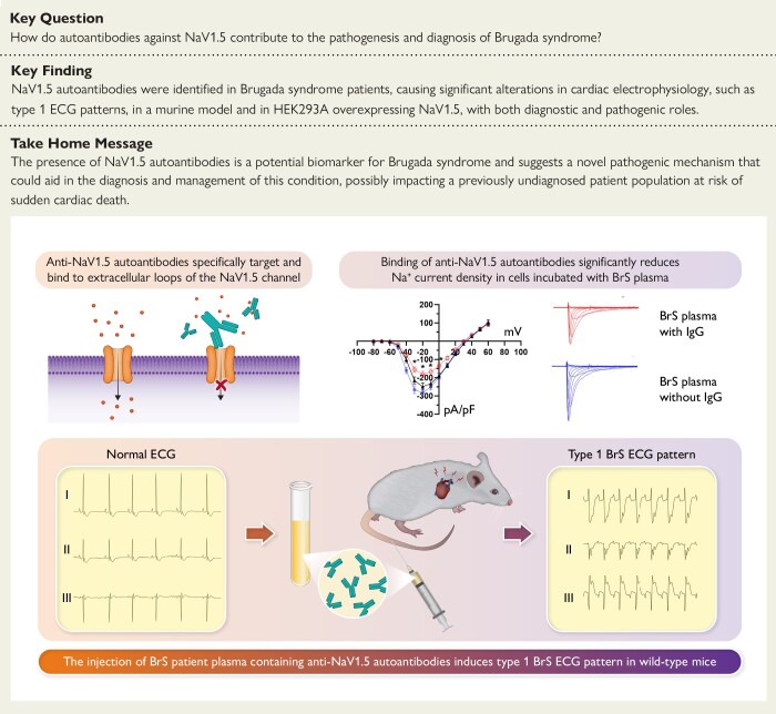
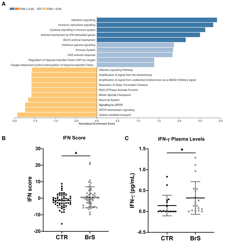
Background and aims: Patients suffering from Brugada syndrome (BrS) are predisposed to life-threatening cardiac arrhythmias. Diagnosis is challenging due to the elusive electrocardiographic (ECG) signature that often requires unconventional ECG lead placement and drug challenges to be detected. Although NaV1.5 sodium channel dysfunction is a recognized pathophysiological mechanism in BrS, only 25% of patients have detectable SCN5A variants. Given the emerging role of autoimmunity in cardiac ion channel function, this study explores the presence and potential impact of anti-NaV1.5 autoantibodies in BrS patients.

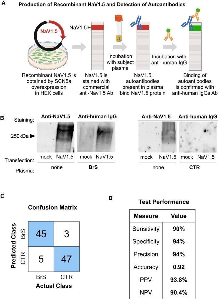
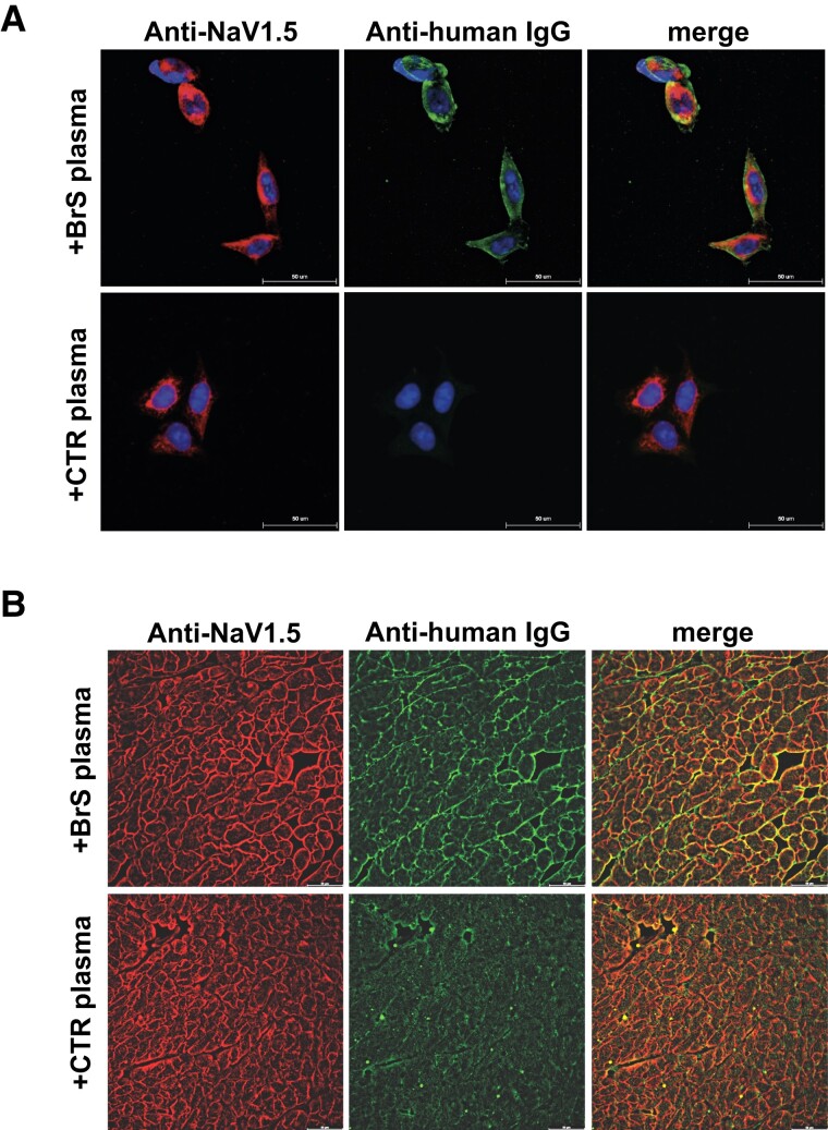
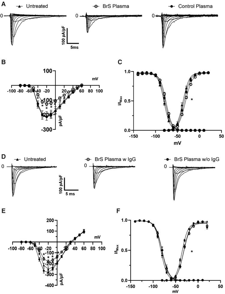
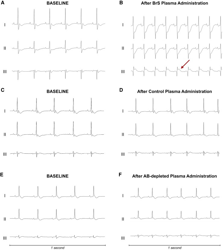
Methods: Using engineered HEK293A cells expressing recombinant NaV1.5 protein, plasma from 50 BrS patients and 50 controls was screened for anti-NaV1.5 autoantibodies via western blot, with specificity confirmed by immunoprecipitation and immunofluorescence. The impact of these autoantibodies on sodium current density and their pathophysiological effects were assessed in cellular models and through plasma injection in wild-type mice.

Results: Anti-NaV1.5 autoantibodies were detected in 90% of BrS patients vs. 6% of controls, yielding a diagnostic area under the curve of .92, with 94% specificity and 90% sensitivity. These findings were consistent across varying patient demographics and independent of SCN5A mutation status. Electrophysiological studies demonstrated a significant reduction specifically in sodium current density. Notably, mice injected with BrS plasma showed Brugada-like ECG abnormalities, supporting the pathogenic role of these autoantibodies.

Conclusions: The study demonstrates the presence of anti-NaV1.5 autoantibodies in the majority of BrS patients, suggesting an immunopathogenic component of the syndrome beyond genetic predispositions. These autoantibodies, which could serve as additional diagnostic markers, also prompt reconsideration of the underlying mechanisms of BrS, as evidenced by their role in inducing the ECG signature of the syndrome in wild-type mice. These findings encourage a more comprehensive diagnostic approach and point to new avenues for therapeutic research.

Keywords: Autoantibodies; Biomarker; Brugada syndrome; NaV1.5.


Figures

Structured Graphical Abstract
Role of anti-NaV1.5 autoantibodies in Brugada syndrome. It demonstrates how these autoantibodies, when present in patient plasma, can elicit the coved-type ST-segment elevation resembling the human type 1 Brugada syndrome electrocardiographic pattern in wild-type mice and significantly reduce sodium current density, supporting their diagnostic and pathogenic significance in the disease. BrS, Brugada syndrome; ECG, electrocardiographic; IgG, immunoglobulin G.
Figure 1
Figure 1
NaV1.5 targeting by plasma immunoglobulin G in Brugada syndrome. (A) Schematic of the workflow for detecting anti-NaV1.5 autoantibodies in Brugada syndrome. HEK293A cells over-expressing NaV1.5 are lysed, and the proteins are resolved via SDS-PAGE. The processed membrane is first probed with a commercial anti-NaV1.5 antibody, followed by incubation with plasma from subjects. Binding of plasma autoantibodies to NaV1.5 is then detected with a secondary anti-human immunoglobulin G antibody. Created with BioRender.com; (B) representative western blots. Left panel: anti-NaV1.5 antibody reveals the presence of the NaV1.5 protein in lysates from NaV1.5-transfected HEK293A cells but not in mock-transfected cells. Right panel: subsequent incubation with Brugada syndrome patient plasma (Brugada syndrome) and control plasma demonstrates specific binding of Brugada syndrome-derived autoantibodies to the NaV1.5 protein, as indicated by the anti-human immunoglobulin G antibody staining, absent in control plasma; (C) confusion matrix for the classification of BrS and control samples based on immunoglobulin G binding to NaV1.5; (D) table summarizing the test performance metrics derived from the confusion matrix in (C) measures of diagnostic efficacy, including sensitivity, specificity, precision, accuracy, and positive and negative predictive values, quantify the reliability of immunoglobulin G binding as a diagnostic marker for Brugada syndrome. BrS, Brugada syndrome; CTR, control; IgG, immunoglobulin G; NPV, negative predictive value; PPV, positive predictive value
Figure 2
Figure 2
Validation of anti-NaV1.5 autoantibody specificity and diagnostic efficacy in Brugada syndrome. (A) Immunofluorescence assays on HEK293A cells over-expressing NaV1.5. Top: Cells treated with plasma from Brugada syndrome patients show specific binding of immunoglobulin Gs (green) to the NaV1.5 channel (red). Bottom: No immunoglobulin G binding is observed in cells treated with control plasma, indicating specificity of the antibody–channel interaction; (B) tissue immunofluorescence demonstrating NaV1.5 channel localization in mouse heart sections. Top: Treatment with Brugada syndrome patient plasma (+Brugada syndrome plasma) results in the co-localization of human immunoglobulin Gs (green) with NaV1.5 expression (red), indicating autoantibody binding to the channel. Bottom: Control plasma (+control plasma) shows no such co-localization, underscoring the specificity of the Brugada syndrome patient autoantibodies. BrS, Brugada syndrome; CTR, control; IgG, immunoglobulin G
Figure 3
Figure 3
Impact of Brugada syndrome patient autoantibodies on NaV1.5 sodium current and channel localization. (A) Representative traces of sodium currents recorded from HEK293A cells over-expressing NaV1.5 untreated (left) or after incubation with Brugada syndrome patient plasma (centre) vs. control plasma (right). The Brugada syndrome panel demonstrates reduced current amplitudes compared with the control panel; (B) current–voltage relationship (I–V curve) of NaV1.5 currents. Data from Brugada syndrome plasma- (n = 70, N = 6, empty circles) and control plasma-treated cells (n = 91, N = 8, filled circles) reveal a marked decrease in peak sodium current in the presence of Brugada syndrome plasma, indicating functional modulation of the channel; untreated cells are in black triangle (n = 35, N = 7); (C) normalized conductance–voltage relationships highlight a shift in the voltage dependence of activation, with Brugada syndrome plasma-treated cells (filled circles) displaying altered channel kinetics compared with controls (empty circles) and untreated cells (filled triangles); (D) families of sodium current elicited in untreated cells (left) or in cells incubated with complete Brugada syndrome plasma (centre) or with BrS plasma devoid of immunoglobulin G (right); (E) I–V curve of the three condition tested: untreated (filled triangles, N = 3, n = 24), Brugada syndrome complete plasma (empty circles, N = 3, n = 25), and Brugada syndrome plasma devoid of immunoglobulin G (black circles, N = 3, n = 36); (F) voltage dependence of channel activation and inactivation (*P < .5). For values, see Supplementary data. BrS, Brugada syndrome; IgG, immunoglobulin G
Figure 4
Figure 4
Electrocardiographic response to plasma in vivo. (A, B) The electrocardiographic recordings from a mouse before (A) and after (B) receiving plasma from a patient with Brugada syndrome. Following the administration, the electrocardiographic mirrors the Brugada-like electrocardiographic pattern commonly observed in humans in bipolar leads, indicated by ST-segment elevation in Lead III and specular ST-segment depression in Leads I and II. This mouse died after the experiment and all the electrocardiographic changes developing after the infusion are shown in detail in Supplementary data. (C, D) The electrocardiographic outcomes from a mouse treated with plasma from a control subject without Brugada syndrome. Notably, no electrocardiographic changes are evident following the infusion (D), indicating the absence of arrhythmogenic activity. (E, F) Electrocardiographic tracings from a mouse receiving antibody-depleted Brugada syndrome plasma. No electrocardiographic abnormalities could be demonstrated following the infusion. Three bipolar lead configurations for electrocardiographic recording are shown (Leads I, II, and III, respectively). Further electrocardiographic details are shown in Supplementary data. AB, antibody; BrS, Brugada syndrome
Figure 5
Figure 5
Transcriptomic and immunologic changes in Brugada syndrome patients. (A) Gene Set Enrichment Analysis: Transcriptomic analysis of PBMCs of Brugada syndrome patients compared with healthy controls shows 540 differentially expressed genes. Gene Set Enrichment Analysis shows significant up-regulation of immune signalling pathways, especially those related to cytokine and interferon signalling. (B) Interferon score: A composite interferon score, calculated as the sum of Z-scores for nine major interferon-related genes, is significantly elevated in Brugada syndrome patients, indicating increased activity of the interferon signalling pathway. (C) Interferon-γ plasma concentrations: Measurement of plasma levels of interferon-γ by enzyme-linked immunosorbent assay further confirms this up-regulation, showing significantly higher interferon-γ levels in Brugada syndrome patients compared with controls. Data were analysed with student t-test (*P < .05). BrS, Brugada syndrome; CTR, control; IFN, interferon

References

    1. Brugada P, Brugada J. Right bundle branch block, persistent ST segment elevation and sudden cardiac death: a distinct clinical and electrocardiographic syndrome. A multicenter report. J Am Coll Cardiol 1992;20:1391–6. 10.1016/0735-1097(92)90253-j - DOI - PubMed
    1. Zeppenfeld K, Tfelt-Hansen J, de Riva M, Winkel BG, Behr ER, Blom NA, et al. . 2022 ESC guidelines for the management of patients with ventricular arrhythmias and the prevention of sudden cardiac death. Eur Heart J 2022;43:3997–4126. 10.1093/eurheartj/ehac262 - DOI - PubMed
    1. Delise P, Allocca G, Sitta N, Migliore F, Dagradi F, Spazzolini C, et al. . Cardiac arrest and Brugada syndrome: is drug-induced type 1 ECG pattern always a marker of low risk? Int J Cardiol 2018;254:142–5. 10.1016/j.ijcard.2017.10.118 - DOI - PubMed
    1. Milman A, Hochstadt A, Andorin A, Gourraud JB, Sacher F, Mabo P, et al. . Time-to-first appropriate shock in patients implanted prophylactically with an implantable cardioverter-defibrillator: data from the Survey on Arrhythmic Events in BRUgada Syndrome (SABRUS). Europace 2019;21:796–802. 10.1093/europace/euy301 - DOI - PubMed
    1. Miyazaki T, Mitamura H, Miyoshi S, Soejima K, Aizawa Y, Ogawa S. Autonomic and antiarrhythmic drug modulation of ST segment elevation in patients with Brugada syndrome. J Am Coll Cardiol 1996;27:1061–70. 10.1016/0735-1097(95)00613-3 - DOI - PubMed

Substances