. 2024 Jul 30;81(1):322.
doi: 10.1007/s00018-024-05366-1.

TMEM9 activates Rab9-dependent alternative autophagy through interaction with Beclin1

Affiliations

TMEM9 activates Rab9-dependent alternative autophagy through interaction with Beclin1

Sohyeon Baek et al. Cell Mol Life Sci. .

Abstract

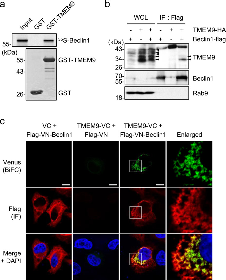
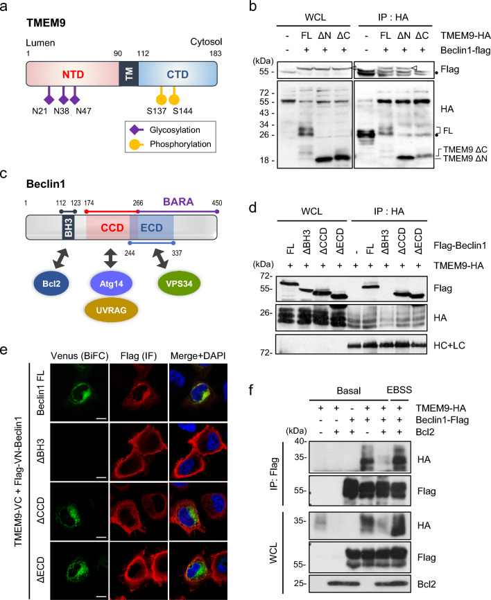
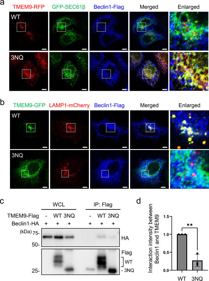
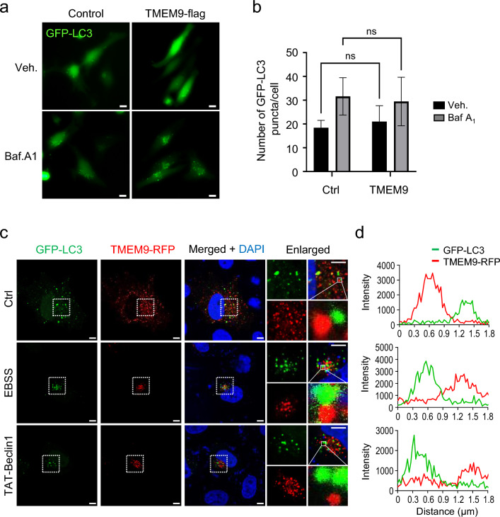
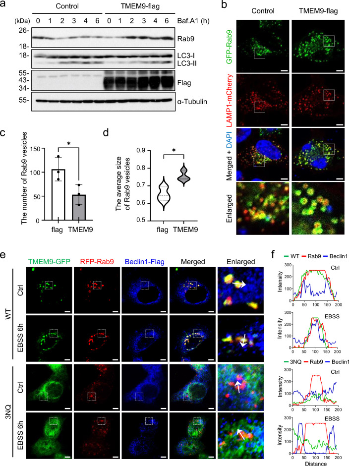
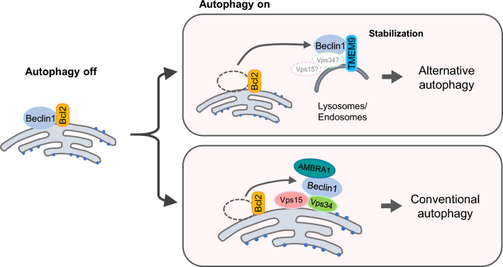
Transmembrane protein 9 (TMEM9) is a transmembrane protein that regulates lysosomal acidification by interacting with the v-type ATPase complex. However, the role of TMEM9 in the lysosome-dependent autophagy machinery has yet to be identified. In this study, we demonstrate that the lysosomal protein TMEM9, which is involved in vesicle acidification, regulates Rab9-dependent alternative autophagy through its interaction with Beclin1. The cytosolic domain of TMEM9 interacts with Beclin1 via its Bcl-2-binding domain. This interaction between TMEM9 and Beclin1 dissociates Bcl-2, an autophagy-inhibiting partner, from Beclin1, thereby activating LC3-independent and Rab9-dependent alternative autophagy. Late endosomal and lysosomal TMEM9 apparently colocalizes with Rab9 but not with LC3. Furthermore, we show that multiple glycosylation of TMEM9, essential for lysosomal localization, is essential for its interaction with Beclin1 and the activation of Rab9-dependent alternative autophagy. These findings reveal that TMEM9 recruits and activates the Beclin1 complex at the site of Rab9-dependent autophagosome to induce alternative autophagy.

Keywords: Alternative autophagy; Beclin1; Rab9; TMEM9.


Conflict of interest statement

The authors declare no conflicting interests.

Figures

Fig. 1
Fig. 1
Lysosomal protein TMEM9 interacts with Beclin1. a GST and GST-TMEM9 proteins were purified by glutathione (GST)-agarose beads and incubated with the 35S-labeled in vitro translated Beclin1. After pulled-down with GST-agarose beads, the reaction products were separated by SDS-PAGE and autoradiography. Input indicates 10% of the in vitro translated Beclin1 protein. b HEK293T cells were transfected with TMEM9-HA and Beclin1-flag as indicated and subjected to immunoprecipitation (IP) assay using Flag-M2 magnetic beads. Whole-cell lysates (WCL) and the immunoprecipitates were analyzed by western blotting using antibodies against TMEM9, Beclin1, and Rab9. c HeLa cells were cotransfected with the VN, VC, TMEM9-VC, and VN-Beclin1 as indicated, and the BiFC assay was monitored under a confocal microscope. Scale bar: 10 μm
Fig. 2
Fig. 2
TMEM9 interacts with Beclin1 through its C terminus domain in a competitive manner with Bcl-2. a TMEM9 domains and the predicted sites of posttranslational modifications, including glycosylation and phosphorylation. NTD, N-terminal domain; CTD, C-terminal domain. b HEK293T cells were co-transfected with Beclin1-flag and either HA-tagged TMEM9 or its deletion mutants (ΔN; 90-183 residues, ΔC;1-90 residues) for 48 h. Cell lysates were immunoprecipitated (IP) using an anti-HA antibody and analyzed by western blotting using anti-flag and anti-HA antibodies. c Schematic representation of Beclin1 domains and its identified interacting partners. d HEK293T cells were co-transfected with TMEM9-HA and full-length (FL) or deletion mutants (∆) of Beclin1-flag. Cell lysates were immunoprecipitated (IP) as indicated in b. e HeLa cells were cotransfected with TMEM9-VC and VN-Beclin1 (FL) or its deletion mutations and visualized under a confocal microscope. Scale bar: 10 μm. f HEK293T cells were co-transfected with TMEM9-HA, Beclin1-flag, and Bcl-2 as indicated and incubated in basal medium or EBSS medium for 6 h. Cell lysates were immunoprecipitated (IP) using an anti-FLAG antibody and analyzed by western blotting using anti-flag, anti-HA, and anti-Bcl2 antibodies
Fig. 3
Fig. 3
The glycosylation of TMEM9 is essential for its lysosomal localization and interaction with Beclin1. a and b HeLa cells were co-transfected with Beclin1-Flag, TMEM9-RFP (WT or 3NQ) (a) or TMEM9-GFP (WT or 3NQ) (b) and either GFP-SEC61β (a) or LAMP1-mCherry (b) for 12 h. Cells were immunostained with an anti-Flag antibody and observed under a confocal microscope. Scale Bar, 5 μm. c and d HEK293T cells were co-transfected with Beclin1-HA and either TMEM9-WT-flag or TMEM9-3NQ-flag. Cell lysates were immunoprecipitated (IP) using Flag-M2 magnetic beads and were analyzed by western blotting using anti-HA and anti-flag antibodies (c). The signals of TMEM9 and Beclin1 on the blots were quantified with ImageJ (d). The data were mean ± s.d. of three independent experiments. **p < 0.01
Fig. 4
Fig. 4
Ectopic expression of TMEM9 does not induce LC-dependent conventional autophagy. a and b HeLa cells were co-transfected with GFP-LC3 and TMEM9-flag for 24 h, treated with 20 nM bafilomycin A1 (Baf.A1) for 6 h, and then observed under the fluorescent microscope. Scale bar, 50 μm (a). The numbers of LC3 dots per cell on images in (a) were counted in (b). c and d HeLa cells were co-transfected with GFP-LC3 and TMEM9-RFP for 24 h, incubated with basal medium, EBSS medium, or 10 μM TAT-Beclin1 for 6 h, and observed under confocal microscope (c). Scale Bar, 5 μm. Fluorescent intensities of GFP (green) and RFP (red) on the indicated area in enlarged images of (c) were measured with ZEISS ZEN software (d)
Fig. 5
Fig. 5
TMEM9 activates Rab9-dependent alternative autophagy. a HeLa cells were transfected with control or TMEM9-flag for 24 h, treated with 20 nM of bafilomycin A1 (Baf.A1) for the indicated times and analyzed with western blotting using anti-Rab9, anti-LC3, anti-flag, and anti-αTubulin antibodies. bd HeLa cells were co-transfected with TMEM9-flag, GFP-Rab9, and LAMP1-mCherry in an indicated manner for 24 h and observed under the confocal microscope (b). The numbers (c) and the sizes (d) of Rab9-positive vesicles on the images were analyzed. The data were mean ± s.d. of three independent experiments. *p < 0.05. Scale Bar, 5 μm. e and f HeLa cells were co-transfected with RFP-Rab9, Beclin1-Flag, and either TMEM9-WT-GFP or TMEM9-3NQ-GFP for 27 h, incubated in control basal (Ctrl) or EBSS medium for 6 h. Cells were immunostained with and anti-Flag antibody and observed under a confocal microscope (e). Scale Bar, 5 μm. Fluorescent intensities of GFP (green), RFP (red), and Alexa Fluor™ Plus 405 (Blue) on the images are measured with ZEISS ZEN software (f)
Fig. 6
Fig. 6
The proposed model shows the role of the TMEM9-Beclin axis in Rab9-dependent alternative autophagy. Under autophagy-inhibitory conditions, Beclin1 interacts with Bcl-2. Upon activation, TMEM9 on the endo/lysosomes replaces Bcl-2 in the Beclin1 complexes, leading to the activation of alternative autophagy utilizing Rab9

References

    1. Mizushima N, Ohsumi Y, Yoshimori T (2002) Autophagosome formation in mammalian cells. Cell Struct Funct 27:421–429. 10.1247/csf.27.421 10.1247/csf.27.421 - DOI - PubMed
    1. Nah J (2023) The role of alternative mitophagy in heart disease. Int J Mol Sci. 10.3390/ijms24076362 10.3390/ijms24076362 - DOI - PMC - PubMed
    1. Pyo JO, Nah J, Jung YK (2012) Molecules and their functions in autophagy. Exp Mol Med 44:73–80. 10.3858/emm.2012.44.2.029 10.3858/emm.2012.44.2.029 - DOI - PMC - PubMed
    1. Nishida Y, Arakawa S, Fujitani K, Yamaguchi H, Mizuta T, Kanaseki T, Komatsu M, Otsu K, Tsujimoto Y, Shimizu S (2009) Discovery of Atg5/Atg7-independent alternative macroautophagy. Nature 461:654–658. 10.1038/nature08455 10.1038/nature08455 - DOI - PubMed
    1. Torii S, Yamaguchi H, Nakanishi A, Arakawa S, Honda S, Moriwaki K, Nakano H, Shimizu S (2020) Identification of a phosphorylation site on Ulk1 required for genotoxic stress-induced alternative autophagy. Nat Commun 11:1754. 10.1038/s41467-020-15577-2 10.1038/s41467-020-15577-2 - DOI - PMC - PubMed

LinkOut - more resources