. 2024 Oct;48(5):3089-3104.
doi: 10.1007/s11259-024-10468-z. Epub 2024 Jul 30.

Genotype classification and pathogenicity of infectious bursal disease virus circulating in vaccinated broiler chicken farms

Affiliations

Genotype classification and pathogenicity of infectious bursal disease virus circulating in vaccinated broiler chicken farms

Samah M Mosad et al. Vet Res Commun. 2024 Oct.

Abstract

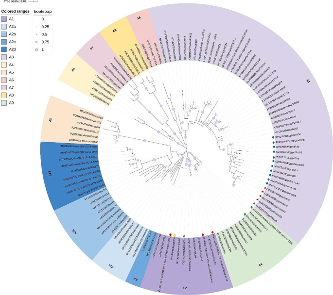
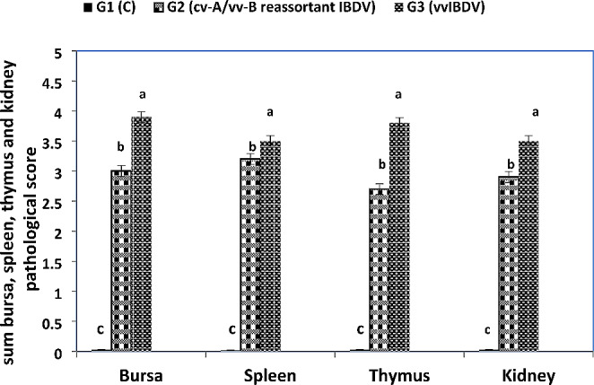
This study investigated the genotype classification and pathogenicity of infectious bursal disease virus (IBDV) circulating in vaccinated broiler chicken farms in Egypt. A total of 150 samples were collected from 30 vaccinated commercial broiler chicken farms and pooled into 30 working samples. IBDV was tested using reverse transcriptase polymerase chain reaction (RT-PCR) amplification of the hypervariable region of the viral protein 2 (hvVP2) and the VP1 gene 5' extremity. Both RT-PCR fragments were sequenced from six samples, and then the obtained nucleotide sequences were analyzed. The IBDV genotypes were identified using nucleotide sequences. Five sequences of the six strains examined were classified as genotype A3B2 for the highly virulent segments A and B (vv-A/vv-B IBDV). Interestingly, this study identified and classified a novel segment-reassortant strain as the A1B2 genotype. Specifically, it involved the segment reassortment of classical virulent segment A (cv-A) with vv-B producing cv-A/vv-B reassortant IBDV. Subsequently, we compared the pathogenicity of reassortant (cv-A/vv-B) IBDV and vvIBDV strains identified in this study. Both strains developed typical IBD clinical signs, postmortem lesions, histopathology, immunohistochemistry, and lesion scores, which were more severe in vvIBDV than reassortant IBDV. In conclusion, this is the first report of the genotype classification based on both genome segments (hvVP2 and VP1) with pathogenicity of IBDV circulating in vaccinated broiler chicken farms and this pathogenicity is more severe in vvIBDV strain than a novel reassortant IBDV strain.

Keywords: Chicken farms; Genotype classification; Pathogenicity; RT-PCR; Reassortment; vvIBDV.


Conflict of interest statement

The authors declare no competing interests.

Figures

Fig. 1
Fig. 1
Phylogenetic tree (Maximum-likelihood) constructed on the nucleotide sequences of hvVP2 gene, which divided into nine major clades (nine genogroups). The nine major genogroups are named A1- A9 according to Gao et al. (2023). Our strains (red triangles) were clustered in genogroup A3 (vvIBDVs) with other Egyptian strains (Blue dots). While the mans1 strain (blue triangle) was grouped in genogroup A1 together with other classic IBDV including the Egyptian “509” classic IBDV strain (yellow dot) and vaccine strains (red dots). Other vaccines were grouped in genogroup A9 together with other attenuated IBDV (green dots)
Fig. 2
Fig. 2
Phylogenetic tree (Maximum-likelihood) established on the nucleotide sequences of VP1 gene, which divided into five major clades (five genogroups). The five major genogroups are named B1- B5 according to Gao et al. (2023). Our strains (red triangles) were clustered in genogroup B2 (vvIBDVs) with other Egyptian strains (yellow dots) in a separate subclade. While all vaccine strains were clustered in genogroup B1 (green dots)
Fig. 3
Fig. 3
BioEdit analysis of deduced amino acid sequences of hvVP2 gene (206- 350 aa) with Giza-Br strain as a reference strain. The alignment revealed the presence of A3 vvIBD amino acid signature in five strains (Red dots). Meanwhile, the mans1 strain (Blue dot) showed the same amino acid profile of A1 classical virulent IBDVs including vaccine strains (green dots). Other vaccine strains A9 (attenuated IBDV) included other vaccine strains (black dots)
Fig. 4
Fig. 4
VP1 gene deduced amino acid sequences (71- 245 aa) analysis by BioEdit software with Egypt 00154 as a reference strain. The alignment revealed the presence of vvIBD amino acid signature in all studied strains (Red dots). Egyptian vvIBDVs have a unique amino acid mutation “V141I”. Vaccine strains (green dots) have non-vvIBDV amino acid signature
Fig. 5
Fig. 5
Embryoic lesions of IBDV isolation in SPF-ECEs. (A) Lesions of cv-A/vv-B IBDV inoculation in 12 days old ECEs 5 days PI: (CE) non-infected control embryo. (IE) cv-A/vv-B IBDV inoculated embryo showing stunted growth, abnormal feathering, and greenish pale liver with pale outlines (red arrow). (B) Lesions of vvIBDV inoculation in 12 days old ECEs 5 days PI: (CE) non-infected control embryo. (IE) embryo inoculated with vvIBDV, showing dwarfism, swollen pale greenish liver (red arrow) with leg deformities (yellow arrows)
Fig. 6
Fig. 6
Postmortem lesions of IBDV experimentally infected chickens. A; B; and C: hemorrhages on thigh and pectoral muscles of chickens infected with cv-A/vv-B reassortant IBDV. D; E and F: severe hemorrhages on thigh muscles of chickens infected with vvIBDV. G: hemorrhages on the proventricular glands in vvIBDV infected chicken. H: vvIBDV infected chicken showing enlarged mottled spleen, enlarged BF (yellow arrow) and nephritis (Red arrow). I: cv-A/vv-B reassortant IBDV infected chicken showing nephritis (Red arrow) and swollen BF with gelatinous exudate (yellow arrow). J: vvIBDV infected chicken showing nephritis (yellow arrow) and swollen hemorrhagic BF with gelatinous exudates
Fig. 7
Fig. 7
Photomicrograph of sections from bursa of Fabricius, spleen, thymus and kidney of chicken infected with IBD stained with H&E X400. (A) Histological picture of bursa of Fabricius shows normal picture in G1 (Control group); (B) normal splenic tissue in G1; (C) Normal cortex and medulla of thymic lobule in G1; (D) Normal renal parenchyma of nephrons; (E) Lymphoid depletion of both cortex and medulla (arrow) and interfollicular fibrous tissue proliferation (arrow head) of bursa in G2; (F) Lymphoid depletion from white and red pulps (arrow) of spleen in G2; (G) Focal area of lymphoid depletion in cortex of thymic lobule (arrow) in G2; (H) Lymphatic aggregates in intersitium (arrow) and necrotic tubules in adjacent parenchyma (arrow head) of kidney in G2; (I) Hyperplastic follicles (arrow) and degenerated epithelia with vesicles formation in covering epithelium (arrow head) of bursa in G3; (J) Hyperplastic white pulps (arrow) of spleen in G3; (K) Mild lymphoid depletion from medulla and replaced by edema (arrow) of thymic lobule in G3; (L) Regenerative attempts from renal tubule epithelia (arrow) and nephrotic tubules in adjacent parenchyma (arrow head) of kidney in G3
Fig. 8
Fig. 8
Immunohistochemistry of experimental infected chickens with IBDV, avidin biotin counter stain with Mayer’s haematoxylin (× 400). (A) Bursa shows moderate positive immunolabeling of IBDV antigen by immunoperoxidase method in the follicular medulla in G2. (B) Bursa shows marked positive immunolabeling of IBDV antigen by immunoperoxidase method in the follicular lymphocytes and interfollicular septa in G3. (C) Spleen shows moderate positive immunolabeling of IBDV antigen in some lymphoid cells in both white and red pulps in G2. (D) Spleen shows marked positive immunolabeling of IBDV antigen in some lymphoid cells in both white and red pulps in G3. (E) Thymus shows positive immunolabeling of IBDV antigen in some lymphocytes in G2. (F) Thymus shows positive immunolabeling of IBDV antigen in some lymphocytes in G3. (G) Kidney shows positive immunolabeling of IBDV antigen in some lining tubular epithelium in G2. (H) Kidney shows marked positive immunolabeling of IBDV antigen in some lining tubular epithelium in G3
Fig. 9
Fig. 9
Statistical analysis of lesions in bursa, spleen, thymus and kidney. C, control; vvIBDV, very virulent IBVD; G1, control; G2, infected with cv-A/vv-B reassortant IBDV; G3, infected with vvIBDV. Statistical analysis showed a higher of total bursa, thymus and kidney lesion score in vvIBDV than cv-A/vv-B reassortant IBDV-infected chickens. Different small alphabetical letters mean significant when P < 0.05

References

    1. Akhila J, Sreeja R, Ambily R, Mini M, Sajitha I (2022) Sequence analysis of VP1, VP2 and VP3 genes of infectious bursal disease virus from a field outbreak in Kerala, India. J Vet Anim Sci 53:633–642
    1. Alkie T, Rautenschlein S (2016) Infectious bursal disease virus in poultry: status and future prospects. Vet Med Res Rep 7:9–18 - PMC - PubMed
    1. Almaw G, Olani A, Sombo M, Yalew B (2022) A very virulent infectious bursal disease virus closely related to New York strain isolated from vaccinated small-scale broiler poultry farm in Addis Ababa, Ethiopia. 10.1101/2022.12.09.519830
    1. Arafat N, Eladl AH, Mahgoub H, El-Shafei RA (2017) Effect of infectious bursal disease (IBD) vaccine on Salmonella Enteritidis infected chickens. Vaccine 35:3682–3689 - PubMed
    1. Bancroft JD, Stevens A, Turner DR (1996) Theory and practice of histological techniques, 4th edn. Churchill Livingston, New York, London

MeSH terms

LinkOut - more resources