. 2024 Aug 6;121(32):e2400783121.
doi: 10.1073/pnas.2400783121. Epub 2024 Jul 30.

In utero delivery of targeted ionizable lipid nanoparticles facilitates in vivo gene editing of hematopoietic stem cells

Affiliations

In utero delivery of targeted ionizable lipid nanoparticles facilitates in vivo gene editing of hematopoietic stem cells

Rohan Palanki et al. Proc Natl Acad Sci U S A. .

Abstract

Monogenic blood diseases are among the most common genetic disorders worldwide. These diseases result in significant pediatric and adult morbidity, and some can result in death prior to birth. Novel ex vivo hematopoietic stem cell (HSC) gene editing therapies hold tremendous promise to alter the therapeutic landscape but are not without potential limitations. In vivo gene editing therapies offer a potentially safer and more accessible treatment for these diseases but are hindered by a lack of delivery vectors targeting HSCs, which reside in the difficult-to-access bone marrow niche. Here, we propose that this biological barrier can be overcome by taking advantage of HSC residence in the easily accessible liver during fetal development. To facilitate the delivery of gene editing cargo to fetal HSCs, we developed an ionizable lipid nanoparticle (LNP) platform targeting the CD45 receptor on the surface of HSCs. After validating that targeted LNPs improved messenger ribonucleic acid (mRNA) delivery to hematopoietic lineage cells via a CD45-specific mechanism in vitro, we demonstrated that this platform mediated safe, potent, and long-term gene modulation of HSCs in vivo in multiple mouse models. We further optimized this LNP platform in vitro to encapsulate and deliver CRISPR-based nucleic acid cargos. Finally, we showed that optimized and targeted LNPs enhanced gene editing at a proof-of-concept locus in fetal HSCs after a single in utero intravenous injection. By targeting HSCs in vivo during fetal development, our Systematically optimized Targeted Editing Machinery (STEM) LNPs may provide a translatable strategy to treat monogenic blood diseases before birth.

Keywords: CRISPR; congenital disease; hematopoietic stem cell; lipid nanoparticles; mRNA.


Conflict of interest statement

Competing interests statement:R.P., M.J.M., and W.H.P. are inventors on a U.S. Provisional Patent Application (No. 63/623,674) related to the technology described in the manuscript.

Figures
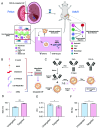
Fig. 1.
Fig. 1.
Design and characterization of CD45R-targeted LNPs. (A) Schematic describing the overall experimental rationale for this study. In brief, LNPs conjugated to CD45 antibody fragments will hone to CD45 receptors expressed on the surface of HSCs within the fetal mouse liver microenvironment. Following LNP internalization in utero and nucleic acid cargo-mediated modification, gene-edited HSCs will undergo normal physiological migration to the bone marrow, where they will engraft and coordinate hematopoiesis of myeloid and lymphoid progeny that also possess the progenitor cell edit. (B) Untargeted LNP formulation scheme that involves C14-490 ionizable lipid, DOPE, cholesterol, C14-PEG2K, and a C18-PEG2K-maleimide (mal-PEG) linker in the organic phase mixed via a microfluidic device with designated RNA cargo in citric acid buffer. (C) Schematic visualizing CD45 F(ab’)2 antibody generation and conjugation to untargeted LNPs to generate targeted LNPs. (D) Characterizing the size, (E) polydispersity (PDI), and (F) encapsulation efficiency of untargeted LNPs (blue) and targeted LNPs (pink). Unpaired parametric Student’s t test (P < 0.05) was used to compare the physiochemical properties of untargeted and targeted LNPs (ns = non-significant, ***P < 0.001); all data reported as the mean ± SEM (minimum n = 3).
Fig. 2.
Fig. 2.
Investigating the efficacy, safety, and mechanism of CD45R-targeted LNPs. (A) Percentage of Jurkat cells (CD45+) expressing GFP 24 h after treatment with untargeted LNPs (blue) or targeted LNPs (pink) encapsulating GFP mRNA at a dose of 100 ng/30,000 cells, visualized via histogram (Left) and plotted (Right). PBS treatment (gray) was used as a negative control. (B) Viability of Jurkat cells following treatment used in (A). (C) Effect of dose (per 30,000 cells) on fold improvement in mRNA delivery to Jurkat (targeted LNP/untargeted LNP). (D) Effect of antibody substitution (CD45 → IgG isotype control) on targeted LNP efficacy in mRNA delivery to Jurkat. (E) Effect of CD45 antibody or (F) IgG antibody pre-treatment on the fold improvement in mRNA delivery to Jurkat (targeted LNP/untargeted LNP). (G) Percentage of HepG2 cells (CD45-) expressing GFP 24 h after treatment with untargeted LNPs or targeted LNPs encapsulating GFP mRNA at a dose of 25 ng/30,000 cells. (H) Volcano plot summarizing the differentially abundant proteins within the corona of plasma-incubated untargeted LNPs and targeted LNPs. (I) Percentage of Jurkat cells expressing GFP after treatment via the approach used in (A) with (P) or without (NP) pre-incubation in plasma. One-way ANOVA with post hoc Dunnett’s test was used to compare the effect of untargeted LNP or targeted LNP treatment in vitro (AC, F, G, and I). Unpaired parametric Student’s t test (P < 0.05) was used to compare the efficacy of CD45 and IgG-targeted LNPs (D) (ns = non-significant, *P < 0.05, **P < 0.01, ***P < 0.001, ****P < 0.0001); all data reported as the mean ± SEM (minimum n = 3).
Fig. 3.
Fig. 3.
Efficacy and safety of targeted LNPs in utero. (A) Percentage of hepatocytes (CD45−/CD31−) or HSCs (Lin−/Sca1+/cKit+) expressing GFP, 60 h after E13.5 R26mT/mG fetal mice were treated with either untargeted LNPs (blue) or targeted LNPs (pink) encapsulating Cre mRNA at a dose of 1 mg/kg. (B) Histological representation of an E14.5 Balb/c mouse fetus (Left) and corresponding fetal livers (Right) from mice treated via in utero IV injection with untargeted LNPs or targeted LNPs encapsulating mCherry mRNA at a dose of 1 mg/kg. Colocalization of CD45 (green) and RFP (red) indicates delivery to CD45+ cells. (C) Percentage of GFP+ hepatocytes and HSCs, 60 h after 12-wk-old adult R26mT/mG mice were treated as described in (A). (D) Percentage of GFP+ hepatocytes and bone marrow HSCs, 4 mo after E13.5 R26mT/mG fetal mice were treated as described in (A). (E) Percentage of myeloid or lymphoid cells in recipient mice expressing GFP at terminal harvest of R26mT/mG mice treated in utero. (F) Percentage of GFP+ hepatocytes and bone marrow HSCs, 4 mo after 12-wk-old adult R26mT/mG mice were treated as described in (A). (G) R26mT/mG fetus survival to birth following in utero IV injection of PBS, untargeted LNPs, or targeted LNPs. (H) Serum aspartate transaminase (AST), (I) alanine transaminase, (J) alkaline phosphatase, and (K) serum cytokine levels of R26mT/mG fetal mice treated at E13.5 with PBS, untargeted LNPs, or targeted LNPs and harvested after 24 h. One-way ANOVA with post hoc Dunnett’s test was used to compare efficacy (A, C, D, and F) and safety (G, H, I, and J) of untargeted and targeted LNPs. Two-way ANOVA with post hoc Šídák’s multiple comparisons test was used to compare the effect of untargeted and targeted LNP treatment on multilineage hematopoietic cell transfection (E) and induction of acute cytokine response (K) (ns = non-significant, *P < 0.05, **P < 0.01, ***P < 0.001, ****P < 0.0001); all data reported as the mean ± SEM (minimum n = 3).
Fig. 4.
Fig. 4.
Secondary transplant of HSCs modified via targeted LNPs in utero. (A) Schematic of secondary transplant study. E13.5 R26mT/mG fetuses were treated with targeted LNPs encapsulating Cre mRNA via in utero IV injection at a dose of 1 mg/kg and followed for 4 mo prior to WBM isolation (donors) and secondary transplanted into lethally irradiated wild-type adult mice (recipients). Recipient mice were subsequently followed for 4 mo via peripheral blood draw prior to terminal BM harvest and assessment of gene modulation in engrafted HSCs. (B) Percentage of CD45+ cells in recipient mice expressing GFP at a given month following secondary transplant. (C) Percentage of myeloid or lymphoid cells in recipient mice expressing GFP at terminal harvest after 4 mo. (D) Percentage of HSCs (Lin−/Sca1+/cKit+) expressing GFP within the bone marrow of donor mice (4 mo after treatment in utero with targeted LNPs) and the bone marrow of recipient mice (4 mo after secondary transplant). One-way ANOVA with post hoc Dunnett’s test was used to compare the level of GFP positivity in recipient mouse CD45+ cells over time (B) (ns = non-significant); all data reported as the mean ± SEM (minimum n = 3).
Fig. 5.
Fig. 5.
Optimization and delivery of gene editing cargo to HSCs in utero. (A) Schematic of DOE approach depicting the design space (4 × 4 × 4 × 4) and sequential library generation (Library A → Library B). (B) Screening LNPs from Library A encapsulating Cas9 mRNA and GFP sgRNA in HepG2-GFP cells at a dose of 100 ng/30,000 cells. Flow cytometry was used to capture resultant GFP knockout after 5 d. Data normalized to the standard formulation (A0, dotted line). (C) Screening LNPs from Library B via the same approach used in (B). (D) Cross-validating B5 LNP formulation encapsulating Cas9 mRNA and GFP sgRNA in Jurkat-GFP (hematopoietic lineage cells) at a dose of 100 ng/30,000 cells. Flow cytometry was used to capture resultant GFP knockout after 5 d. (E) Testing the gene editing efficacy of unoptimized (A0) and optimized (B5) LNPs encapsulating Cas9 mRNA and TTR sgRNA after in utero IV administration at a dose of 1 mg/kg into E13.5 wild-type mice. Genomic DNA from the fetal liver was harvested after 5 d, and indels at the intended locus were quantified via NGS. PBS-injected fetuses were used as a negative control. (F) Schematic of experiment evaluating the gene editing efficacy of optimized untargeted LNPs (B5) and optimized targeted LNPs (STEM) via the same approach used in (E), although HSCs (Lin−/Sca1+/cKit+) were also isolated and sequenced. (G) Indels at the intended locus in genomic DNA from the fetal liver of animals treated with PBS, B5 LNPs, or STEM LNPs. (H) Indels at the intended locus in genomic DNA from HSCs of animals treated with PBS, B5 LNPs, or STEM LNPs. One-way ANOVA with post hoc Dunnett’s test was used to compare gene editing efficacy of LNP formulations (BE, G, and H) (ns = non-significant, *P < 0.05, **P < 0.01, ***P < 0.001, ****P < 0.0001); all data reported as the mean ± SEM (minimum n = 3).

References

    1. Kohne E., Hemoglobinopathies. Dtsch. Arztebl. Int. 108, 532–540 (2011). - PMC - PubMed
    1. Harteveld C. L., et al. , The hemoglobinopathies, molecular disease mechanisms and diagnostics. Int. J. Lab. Hematol. 44, 28–36 (2022). - PMC - PubMed
    1. Chui D. H. K., Waye J. S., Hydrops fetalis caused by α-thalassemia: An emerging health care problem. Blood 91, 2213–2222 (1998). - PubMed
    1. Palanki R., Peranteau W. H., Mitchell M. J., Delivery technologies for in utero gene therapy. Adv. Drug Deliv. Rev. 169, 51 (2021). - PMC - PubMed
    1. Prakash V., Moore M., Yáñez-Muñoz R. J., Current progress in therapeutic gene editing for monogenic diseases. Mol. Ther. 24, 465–474 (2016). - PMC - PubMed