. 2024 Aug 1;72(8):1124-1129.
doi: 10.4103/IJO.IJO_3108_23. Epub 2024 Jul 29.

Outcomes of keratoplasty in a cohort of Pythium insidiosum keratitis cases at a tertiary eye care center in India

Affiliations

Outcomes of keratoplasty in a cohort of Pythium insidiosum keratitis cases at a tertiary eye care center in India

Manisha Acharya et al. Indian J Ophthalmol. .

Abstract

Purpose: To assess outcomes of keratoplasty performed in patients diagnosed with keratitis caused by Pythium insidiosum (PI).

Design: Retrospective review.

Methods: Preoperative, intra operative and post operative data of patients diagnosed with PI keratitis and who underwent keratoplasty for their condition from January 2020 to December 2021 were collected from the central patient database of a tertiary eye care hospital in India. The data were analyzed for anatomic success, elimination of infection, graft survival, incidence of repeat keratoplasty, final visual acuity and varied complications.

Results: In total, 16 eyes underwent penetrating keratoplasty for PI keratitis during the study period. Mean time to keratoplasty from onset of symptoms was 31.3 days and mean graft size was 10.4 mm. Nine out of the 16 cases had recurrence of infection following surgery, seven of which required a repeat keratoplasty for elimination of infection. Mean graft size for repeat keratoplasty performed in recurrent cases was 11.7 mm. Globe was successfully salvaged in 14 out of 16 patients (87.5 %). Three grafts remained clear at 6-month follow up while 11 grafts failed. Mean improvement in uncorrected visual acuity from 2.32 to 2.04 logMAR was observed at last follow up. Endo-exudates, graft infiltration, graft dehiscence, secondary glaucoma and retinal detachment were the various complications noted after keratoplasty.

Conclusion: PI keratitis is a tenacious and potentially blinding condition. Keratoplasty remains the choice of treatment in this condition, however recurrence of disease and graft failure are common. Large sized grafts, meticulous per-operative removal of infection, adjuvant cryotherapy, and intraoperative and post operative use of antibiotics can help in improving outcome of keratoplasty in these patients.


Conflict of interest statement

There are no conflicts of interest.

Figures

Figure 1
Figure 1
(a) Clinical photograph of a 58-year-old man with Pythium keratitis with limbal involvement (red arrow). (b) Large graft with complete removal of infection on postoperative day 1. (c) Failed graft with no recurrence seen at a 2-month follow-up. (d) Slit-lamp picture of a 19-year-old girl with mid-stromal tentacle-like lesions (red arrow) characteristic of Pythium keratitis. (e) Progression of disease leading to central corneal perforation with flat anterior chamber despite medical management of Pythium. (f) Postoperative day 4 picture following therapeutic keratoplasty, showing clear graft with no recurrence
Figure 2
Figure 2
(a) Clinical picture of a 21-year-old girl with Pythium keratitis. (b) Graft dehiscence and endoexudates (red arrow) observed on postoperative day 26. (c) Hazy graft with no recurrence of disease seen after 1 month. (d) Slit-lamp examination of a 24-year-old male with Pythium keratitis. (e) Recurrence observed on fourth postoperative day (red arrow). (f) Clear and compact graft with no recurrence of infection at postoperative week 3. (g) Clinical presentation of a 34-year-old man with Pythium keratitis. (h) Recurrence noted at 1 week as infiltration at the graft–host junction and hypopyon (red arrows). (i) Postoperative Day 22 of repeat keratoplasty shows clear and healthy graft
Figure 3
Figure 3
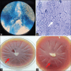
(a and b) Potassium hydroxide with calcofluor white smear showing septate, broad, acute-angled, nonfilamentous hyphae from corneal scraping. (c and d) Blood agar showing radiating colonies with flat planes, feathery edges, partially submerged, glabrous growth suggestive of Pythium insidiosum
Figure 4
Figure 4
Kaplan–Meier survival curve of grafts which developed recurrence of Pythium insidiosum keratitis and underwent repeat keratoplasty. The average time between two surgeries in these cases was estimated to be 47 days (range 11–93 days). Patients with no recurrence of the disease were followed up for 497 days on average

References

    1. Ung L, Bispo PJM, Shanbhag SS, Gilmore MS, Chodosh J. The persistent dilemma of microbial keratitis: Global burden, diagnosis, and antimicrobial resistance. Surv Ophthalmol. 2019;64:255–71. - PMC - PubMed
    1. Whitcher JP, Srinivasan M. Corneal ulceration in the developing world--a silent epidemic. Br J Ophthalmol. 1997;81:622–3. - PMC - PubMed
    1. Van Gelder RN. Ocular pathogens for the twenty-first century. Am J Ophthalmol. 2010;150:595–7. - PMC - PubMed
    1. Hasika R, Lalitha P, Radhakrishnan N, Rameshkumar G, Prajna NV, Srinivasan M. Pythium keratitis in South India: Incidence, clinical profile, management, and treatment recommendation. Indian J Ophthalmol. 2019;67:42–7. - PMC - PubMed
    1. Gaastra W, Lipman LJ, De Cock AW, Exel TK, Pegge RB, Scheurwater J, et al. Pythium insidiosum: An overview. Vet Microbiol. 2010;146:1–16. - PubMed

MeSH terms