Comparative Study
. 2024 Winter;26(2):101069.
doi: 10.1016/j.jocmr.2024.101069. Epub 2024 Jul 28.

Clinical utility of a rapid two-dimensional balanced steady-state free precession sequence with deep learning reconstruction

Affiliations
Comparative Study

Clinical utility of a rapid two-dimensional balanced steady-state free precession sequence with deep learning reconstruction

Katerina Eyre et al. J Cardiovasc Magn Reson. 2024 Winter.

Abstract

Background: Cardiovascular magnetic resonance (CMR) cine imaging is still limited by long acquisition times. This study evaluated the clinical utility of an accelerated two-dimensional (2D) cine sequence with deep learning reconstruction (Sonic DL) to decrease acquisition time without compromising quantitative volumetry or image quality.

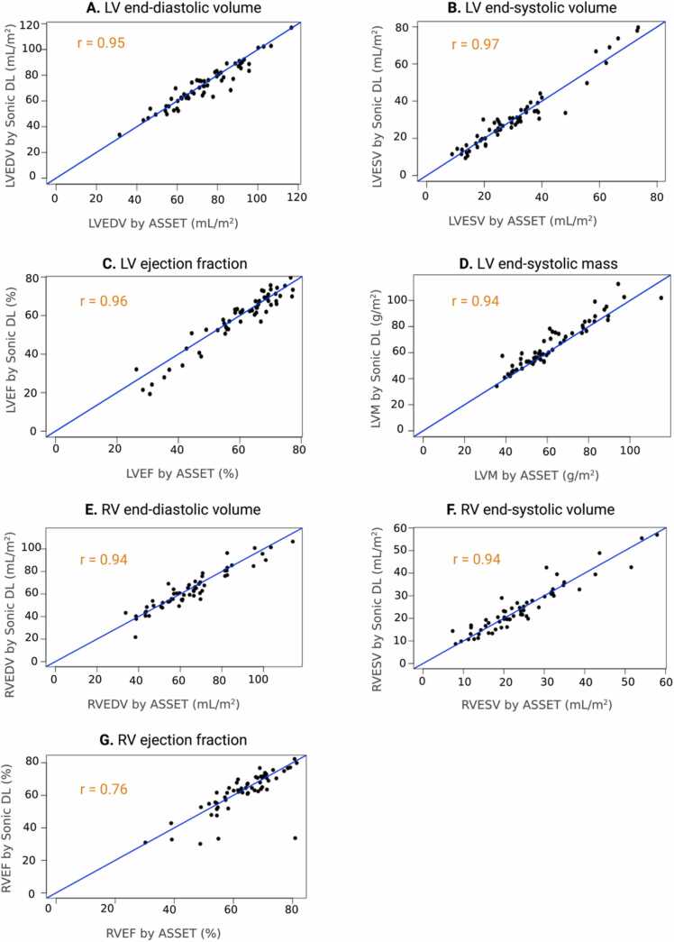
Methods: A sub-study using 16 participants was performed using Sonic DL at two different acceleration factors (8× and 12×). Quantitative left-ventricular volumetry, function, and mass measurements were compared between the two acceleration factors against a standard cine method. Following this sub-study, 108 participants were prospectively recruited and imaged using a standard cine method and the Sonic DL method with the acceleration factor that more closely matched the reference method. Two experienced clinical readers rated images based on their diagnostic utility and performed all image contouring. Quantitative contrast difference and endocardial border sharpness were also assessed. Left- and right-ventricular volumetry, left-ventricular mass, and myocardial strain measurements were compared between cine methods using Bland-Altman plots, Pearson's correlation, and paired t-tests. Comparative analysis of image quality was measured using Wilcoxon-signed-rank tests and visualized using bar graphs.

Results: Sonic DL at an acceleration factor of 8 more closely matched the reference cine method. There were no significant differences found across left ventricular volumetry, function, or mass measurements. In contrast, an acceleration factor of 12 resulted in a 6% (5.51/90.16) reduction of measured ejection fraction when compared to the standard cine method and a 4% (4.32/88.98) reduction of measured ejection fraction when compared to Sonic DL at an acceleration factor of 8. Thus, Sonic DL at an acceleration factor of 8 was chosen for downstream analysis. In the larger cohort, this accelerated cine sequence was successfully performed in all participants and significantly reduced the acquisition time of cine images compared to the standard 2D method (reduction of 37% (5.98/16) p < 0.0001). Diagnostic image quality ratings and quantitative image quality evaluations were statistically not different between the two methods (p > 0.05). Left- and right-ventricular volumetry and circumferential and radial strain were also similar between methods (p > 0.05) but left-ventricular mass and longitudinal strain were over-estimated using the proposed accelerated cine method (mass over-estimated by 3.36 g/m2, p < 0.0001; longitudinal strain over-estimated by 1.97%, p = 0.001).

Conclusion: This study found that an accelerated 2D cine method with DL reconstruction at an acceleration factor of 8 can reduce CMR cine acquisition time by 37% (5.98/16) without significantly affecting volumetry or image quality. Given the increase of scan time efficiency, this undersampled acquisition method using deep learning reconstruction should be considered for routine clinical CMR.

Keywords: Accelerated imaging; CMR; Cardiac function; Clinical utility; Deep learning reconstruction; Diagnostic accuracy.


Conflict of interest statement

Declaration of competing interests The authors declare the following financial interests/personal relationships which may be considered as potential competing interests: Matthias Friedrich reports financial support was provided by MEDTEQ and GE Healthcare. Matthias Friedrich reports a relationship with Circle Cardiovascular Imaging Inc that includes board membership. Martin Janich, Junjie Ma, and Xucheng Zhu report a relationship with GE Healthcare that includes employment. The other authors declare that they have no known competing financial interests or personal relationships that could have appeared to influence the work reported in this paper.

Figures

ga1
Graphical abstract
Fig. 1
Fig. 1
Schematic depiction of the calculation of endocardial edge sharpness measurements. (A) Radial lines drawn in an orthogonal fashion from the center of the LV cavity to the subepicardial myocardial boundary to compute a signal intensity profile. (B) Endocardial edge sharpness was calculated by taking the average slope of the sigmoid functions that were fit to the signal intensity profile. LV left ventricle.
Fig. 2
Fig. 2
Comparison of left-ventricular (LV): (A) end-diastolic volume (EDV), (B) end-systolic volume (ESV), (C) ejection fraction (EF) and (D) mass between ASSET and Sonic DL at an acceleration factor of 8 and 12. A repeated measures ANOVA was used to compare means between methods. A p-value of <0.05 was considered statistically significant. ASSET array spatial sensitivity encoding technique, DL deep learning, n.s. non-significant, LVM left ventricular mass, ANOVA analysis of variance.
Fig. 3
Fig. 3
Comparison of scan time between methods. (A) Comparison of scan time between acquisition methods of the complete short-axis (SAx) and long-axis (LAx) views. (B) Comparison of scan time between methods of the SAx stack. (C) Comparison of scan time between methods of the three LAx views. ASSET array coil spatial sensitivity encoding, bSSFP balanced steady-state free precession, DL deep learning.
Fig. 4
Fig. 4
Results from image quality assessment. (A) Diagnostic confidence scores were obtained from two experienced clinical readers. Images were anonymized to sequence type and randomized with respect to the order they were presented to the readers. 1: No diagnostic confidence (not interpretable); 2: low diagnostic confidence (poor image quality); 3: medium diagnostic confidence (good overall image quality with one or two views with poorer IQ); 4: high diagnostic confidence (perfect image quality). (B) Contrast difference measurements were taken between the blood pool and the myocardium by subtracting the signal intensity of the myocardium from that of the blood pool. (C) Endocardial edge sharpness was calculated by taking the average slope of the sigmoid functions that were fit to the signal intensity profile of the line segment than was drawn orthogonal to the myocardial and blood pool border. ASSET array coil spatial sensitivity encoding technique, bSSFP balanced steady-state free precession, DL deep learning, SI signal intensity, BP blood pool, Myo myocardium, IQ image quality.
Fig. 5
Fig. 5
A representative set of cine images at end-systole and end-diastole from a patient with atrial fibrillation. ASSET array coil spatial sensitivity encoding technique, bSSFP balanced steady-state free precession, DL deep learning, LAx long axis.
Fig. 6
Fig. 6
Bland-Altman plots displaying the similarity of measured ventricular function metrics using ASSET bSSFP and Sonic DL bSSFP sequence. The solid blue line represents the average bias between measurements. The dotted lines represent the upper and lower 95% limits of agreement. Bland-Altman plots displayed for (A) LVEDV, (B) LVESV, (C) LVEF, (D) LVM, (E) RVEDV, (F) RVESV, (G) RVEF. LV: left ventricle, ASSET array coil spatial sensitivity encoding technique encoding, DL deep learning, RV right ventricle, EDV end-diastolic volume, ESV end-systolic volume, EF ejection fraction, LVM left ventricular mass.
Fig. 7
Fig. 7
Scatter plot displaying the relationship between measured ventricular function metrics by ASSET bSSFP and Sonic DL bSSFP. Scatter plots are displayed for (A) LVEDV, (B) LVESV, (C) LVEF, (D) LVM, (E) RVEDV, (F) RVESV, and (G) RVEF. LV left ventricle, ASSET array coil spatial sensitivity encoding technique, DL deep learning, RV right ventricle, EDV end-diastolic volume, ESV end-systolic volume, EF ejection fraction, LVM left ventricular mass, r correlation coefficient.

Similar articles

Cited by

References

    1. Schulz-Menger J., Bluemke D.A., Bremerich J., Flamm S.D., Fogel M.A., Friedrich M.G., et al. Standardized image interpretation and post-processing in cardiovascular magnetic resonance - 2020 update. J Cardiovasc Magn Reson. 2020;22(1):19. doi: 10.1186/s12968-020-00610-6. - DOI - PMC - PubMed
    1. Grothues F., Smith G.C., Moon J.C.C., Bellenger N.G., Collins P., Klein H.U., et al. Comparison of interstudy reproducibility of cardiovascular magnetic resonance with two-dimensional echocardiography in normal subjects and in patients with heart failure or left ventricular hypertrophy. Am J Cardiol. 2002;90(1):29–34. https://www.sciencedirect.com/science/article/pii/S0002914902023810 - PubMed
    1. Kramer C.M., Barkhausen J., Bucciarelli-Ducci C., Flamm S.D., Kim R.J., Nagel E. Standardized cardiovascular magnetic resonance imaging (CMR) protocols: 2020 update. J Cardiovasc Magn Reson. 2020;22(1):17. doi: 10.1186/s12968-020-00607-1. - DOI - PMC - PubMed
    1. Jahnke C., Paetsch I., Achenbach S., Schnackenburg B., Gebker R., Fleck E., et al. Coronary MR imaging: breath-hold capability and patterns, coronary artery rest periods, and β-blocker use. Radiology. 2006;239(1):71–78. doi: 10.1148/radiol.2383042019. - DOI - PubMed
    1. Chandler A.G., Pinder R.J., Netsch T., Schnabel J.A., Hawkes D.J., Hill D.L., et al. Correction of misaligned slices in multi-slice cardiovascular magnetic resonance using slice-to-volume registration. J Cardiovasc Magn Reson. 2008;10(1):13. doi: 10.1186/1532-429X-10-13. - DOI - PMC - PubMed

Publication types

LinkOut - more resources