. 2024 Jul 30;11(7):ENEURO.0035-24.2024.
doi: 10.1523/ENEURO.0035-24.2024. Print 2024 Jul.

Exploring Kainic Acid-Induced Alterations in Circular Tripartite Networks with Advanced Analysis Tools

Affiliations

Exploring Kainic Acid-Induced Alterations in Circular Tripartite Networks with Advanced Analysis Tools

Andrey Vinogradov et al. eNeuro. .

Abstract

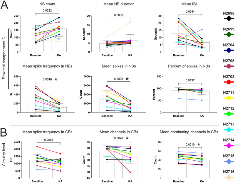
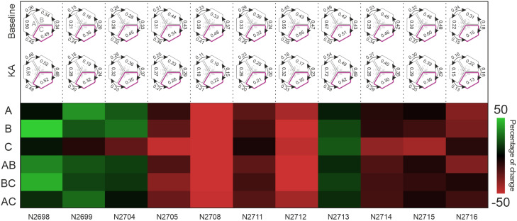
Brain activity implies the orchestrated functioning of interconnected brain regions. Typical in vitro models aim to mimic the brain using single human pluripotent stem cell-derived neuronal networks. However, the field is constantly evolving to model brain functions more accurately through the use of new paradigms, e.g., brain-on-a-chip models with compartmentalized structures and integrated sensors. These methods create novel data requiring more complex analysis approaches. The previously introduced circular tripartite network concept models the connectivity between spatially diverse neuronal structures. The model consists of a microfluidic device allowing axonal connectivity between separated neuronal networks with an embedded microelectrode array to record both local and global electrophysiological activity patterns in the closed circuitry. The existing tools are suboptimal for the analysis of the data produced with this model. Here, we introduce advanced tools for synchronization and functional connectivity assessment. We used our custom-designed analysis to assess the interrelations between the kainic acid (KA)-exposed proximal compartment and its nonexposed distal neighbors before and after KA. Novel multilevel circuitry bursting patterns were detected and analyzed in parallel with the inter- and intracompartmental functional connectivity. The effect of KA on the proximal compartment was captured, and the spread of this effect to the nonexposed distal compartments was revealed. KA induced divergent changes in bursting behaviors, which may be explained by distinct baseline activity and varied intra- and intercompartmental connectivity strengths. The circular tripartite network concept combined with our developed analysis advances importantly both face and construct validity in modeling human epilepsy in vitro.

Keywords: brain-on-a-chip; burst; microelectrode array; neuronal connectivity; neuronal culture; signal processing.


Conflict of interest statement

The authors declare no competing financial interests. The MEMO platform and its associated circuitry burst detection analysis software are currently in the process of commercialization, meaning that part of software elements are under IP evaluation.

Figures

None
Visual Abstract
Figure 1.
Figure 1.
Stages of the development of the method. (1) Pipeline of algorithm evaluation and template selection and (2) iterative feature enhancement process.
Figure 2.
Figure 2.
Network and CB detection pipeline. A, Steps of the novel method. The detected spikes are pooled into a histogram, and the ISIN threshold is defined for initial NB detection. B, The first five steps are visualized with single-compartment raster segments of various lengths. The green and red vertical lines represent the start and end times, respectively, of the captured local NBs. The blue arrows in STEPS 3 and 4 indicate limitations of the simple “minimum participating channels” criterion. C, The last step (STEP 6) is shown as a combined raster of three MEMO compartments. The purple polygons at the top depict time intervals with detected circuitry-level bursts involving all three compartments; the orange polygons indicate intermediate circuitry-level bursts with two-compartment synchronicity; and the yellow polygons indicate local NBs in a single compartment. The length of the polygons corresponds to the duration of the corresponding phenomenon.
Figure 3.
Figure 3.
Cumulative circuitry-level burst duration. The scatter plots depict scores for the derived metric among 5,000 simulations for 11 MEMOs labeled with their MEMO platform identification codes. The simulations included compartmental NB shuffling for each plate for two conditions: baseline and KA exposure. The real observed condition scores are indicated with red triangles. The bold line on each scatter plot represents the mean value for the scores of 5,000 simulations together with the single entity calculated from the observed data.
Figure 4.
Figure 4.
Pairwise comparisons. A, Six of the eight NB output parameters from the proximal C compartment and B, Three of the eight CB output parameters that showed significant (p < 0.05) KA-induced alterations prior to Bonferroni’s adjustment of the significance level. Wilcoxon matched-pairs signed-rank test. The color codes for the corresponding MEMO labels are shown on the side. The bar charts with whiskers present means and standard deviations. n = 11 for each pairwise comparison. The p values are presented on top of each plot. The NB/CB parameters with alterations that remained significant (p < 0.00625) after Bonferroni’s correction for the families of eight hypotheses are marked with asterisk *. The full lists of pairwise comparisons for the NB and CB parameters are available in Extended Data Figures 4-1–4-4.
Figure 5.
Figure 5.
Binned heatmap of baseline-normalized percentage changes in eight parameters in three compartments and at the circuitry level among 11 MEMOs. The last 5fmin bin of the baseline recording was used to calculate the percentage change in the subsequent six 5fmin bins recorded after KA exposure. The upper and lower limits of percentage change were set to 50% for visualization purposes.
Figure 6.
Figure 6.
Functional connectivity analysis results before and after KA exposure. In the upper half of the figure, the average intracompartmental and intercompartmental average connectivity values are plotted for both experimental conditions for all 11 MEMOs. The exposed proximal C compartment is highlighted in purple. The first scheme embeds the compartment labels. The lower half of the figure shows the heatmap of percentage changes in the connectivity values after KA exposure. The upper and lower limits of percentage change were set to 50% for visualization purposes. Extended Data Figure 6-1 shows pairwise comparisons of average intra- and intercompartmental CorSE values before and after KA exposure among 11 MEMOs. Extended Data Figure 6-2 presents pairwise comparisons of average intra- and intercompartmental CorSE values within each MEMO.

Similar articles

References

    1. Alsaqati M, Heine VM, Harwood AJ (2020) Pharmacological intervention to restore connectivity deficits of neuronal networks derived from ASD patient iPSC with a TSC2 mutation. Mol Autism 11:80. 10.1186/s13229-020-00391-w - DOI - PMC - PubMed
    1. Bakkum D, Radivojevic M, Frey U, Franke F, Hierlemann A, Takahashi H (2014) Parameters for burst detection. Front Comput Neurosci 7:193. 10.3389/fncom.2013.00193 - DOI - PMC - PubMed
    1. Bassett DS, Sporns O (2017) Network neuroscience. Nat Neurosci 20:353–364. 10.1038/nn.4502 - DOI - PMC - PubMed
    1. Belitski A, Gretton A, Magri C, Murayama Y, Montemurro MA, Logothetis NK, Panzeri S (2008) Low-frequency local field potentials and spikes in primary visual cortex convey independent visual information. J Neurosci 28:5696–5709. 10.1523/JNEUROSCI.0009-08.2008 - DOI - PMC - PubMed
    1. Brofiga M, Pisano M, Tedesco M, Boccaccio A, Massobrio P (2022) Functional inhibitory connections modulate the electrophysiological activity patterns of cortical–hippocampal ensembles. Cereb Cortex 32:1866–1881. 10.1093/cercor/bhab318 - DOI - PubMed

LinkOut - more resources