. 2024 Dec;67(12):2786-2803.
doi: 10.1007/s00125-024-06232-2. Epub 2024 Jul 30.

Deletion of RFX6 impairs iPSC-derived islet organoid development and survival, with no impact on PDX1+/NKX6.1+ progenitors

Affiliations

Deletion of RFX6 impairs iPSC-derived islet organoid development and survival, with no impact on PDX1+/NKX6.1+ progenitors

Noura Aldous et al. Diabetologia. 2024 Dec.

Abstract

Aims/hypothesis: Homozygous mutations in RFX6 lead to neonatal diabetes accompanied by a hypoplastic pancreas, whereas heterozygous mutations cause MODY. Recent studies have also shown RFX6 variants to be linked with type 2 diabetes. Despite RFX6's known function in islet development, its specific role in diabetes pathogenesis remains unclear. Here, we aimed to understand the mechanisms underlying the impairment of pancreatic islet development and subsequent hypoplasia due to loss-of-function mutations in RFX6.

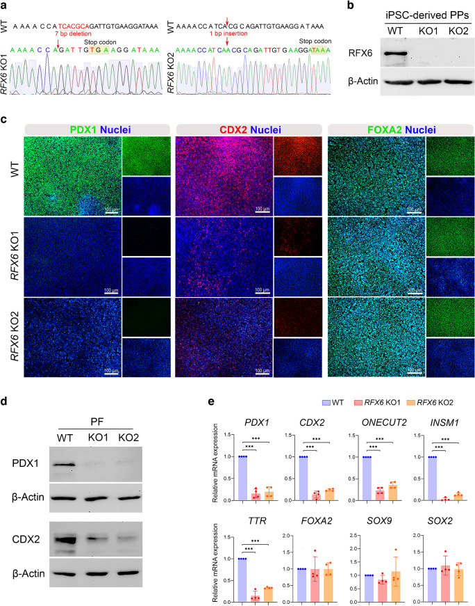
Methods: We examined regulatory factor X6 (RFX6) expression during human embryonic stem cell (hESC) differentiation into pancreatic islets and re-analysed a single-cell RNA-seq dataset to identify RFX6-specific cell populations during islet development. Furthermore, induced pluripotent stem cell (iPSC) lines lacking RFX6 were generated using CRISPR/Cas9. Various approaches were then employed to explore the consequences of RFX6 loss across different developmental stages. Subsequently, we evaluated transcriptional changes resulting from RFX6 loss through RNA-seq of pancreatic progenitors (PPs) and endocrine progenitors (EPs).

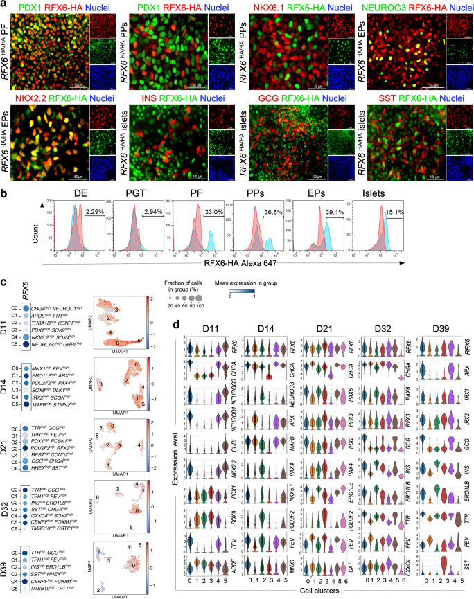
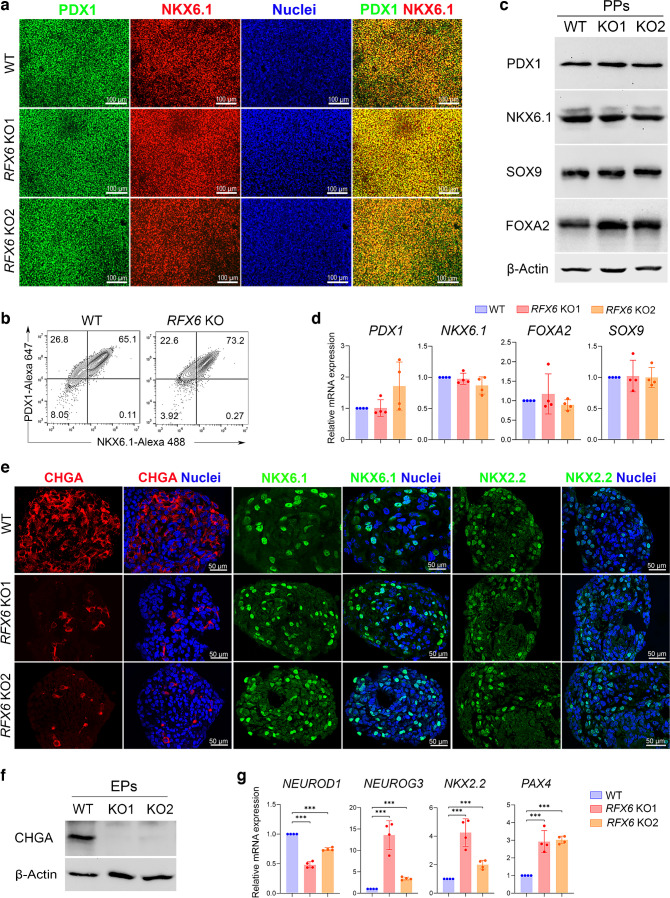
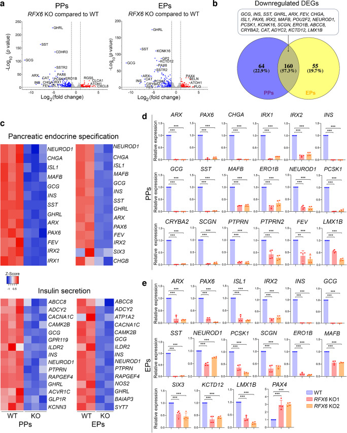
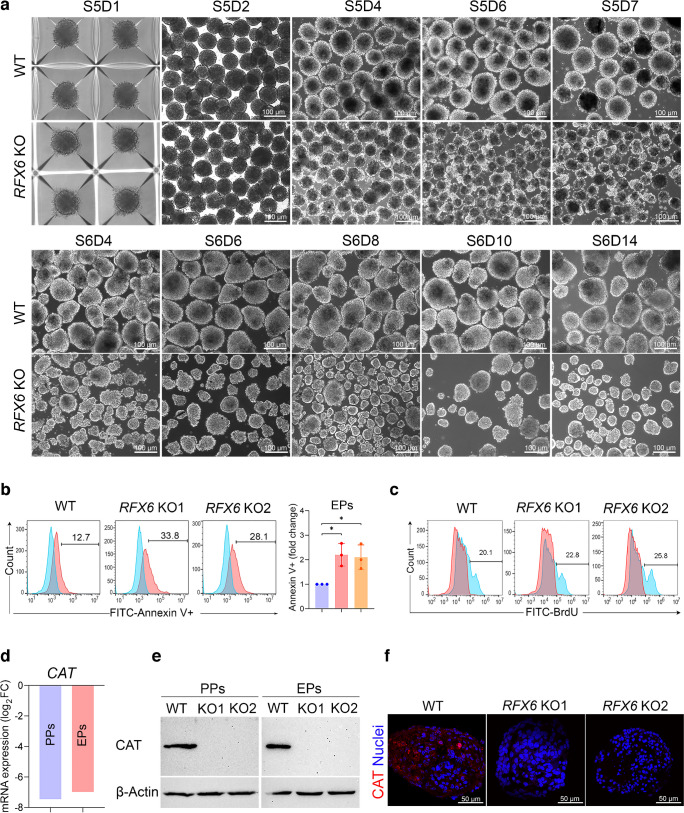
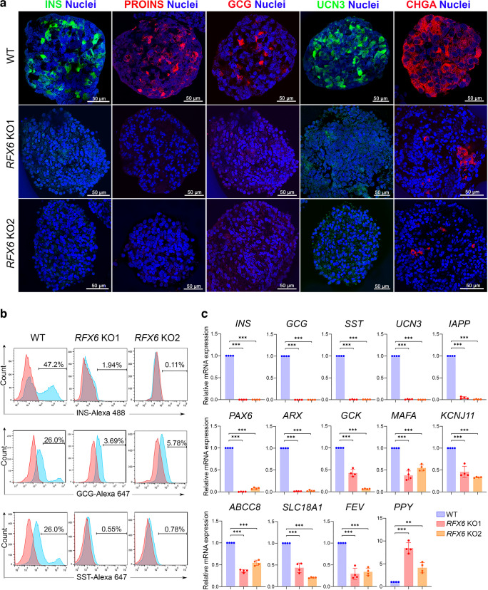
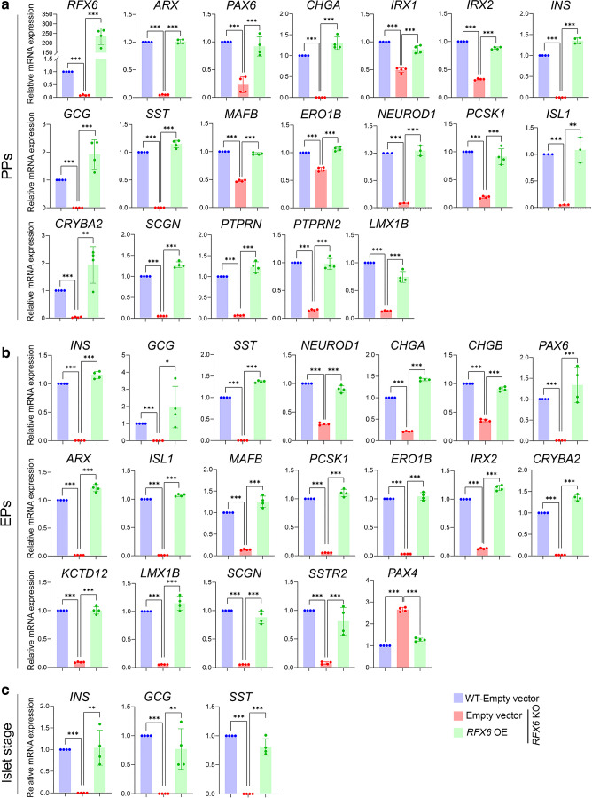
Results: RFX6 expression was detected in PDX1+ cells in the hESC-derived posterior foregut (PF). However, in the PPs, RFX6 did not co-localise with pancreatic and duodenal homeobox 1 (PDX1) or NK homeobox 1 (NKX6.1) but instead co-localised with neurogenin 3, NK2 homeobox 2 and islet hormones in the EPs and islets. Single-cell analysis revealed high RFX6 expression levels in endocrine clusters across various hESC-derived pancreatic differentiation stages. Upon differentiating iPSCs lacking RFX6 into pancreatic islets, a significant decrease in PDX1 expression at the PF stage was observed, although this did not affect PPs co-expressing PDX1 and NKX6.1. RNA-seq analysis showed the downregulation of essential genes involved in pancreatic endocrine differentiation, insulin secretion and ion transport due to RFX6 deficiency. Furthermore, RFX6 deficiency resulted in the formation of smaller islet organoids due to increased cellular apoptosis, linked to reduced catalase expression, implying a protective role for RFX6. Overexpression of RFX6 reversed defective phenotypes in RFX6-knockout PPs, EPs and islets.

Conclusions/interpretation: These findings suggest that pancreatic hypoplasia and reduced islet cell formation associated with RFX6 mutations are not due to alterations in PDX1+/NKX6.1+ PPs but instead result from cellular apoptosis and downregulation of pancreatic endocrine genes.

Data availability: RNA-seq datasets have been deposited in the Zenodo repository with accession link (DOI: https://doi.org/10.5281/zenodo.10656891 ).

Keywords: Diabetes; Endocrine specification; Islet organoids; Pancreatic hypoplasia; Pancreatic progenitors; Transcription factors.


Conflict of interest statement

Acknowledgements: We would like to thank N. R. Dunn (A*STAR, Singapore) for providing the HA-RFX6 tagged H9 hESC lines (RFX6HA/HA H9-hESCs). Furthermore, we thank the Genomic Core members at QBRI for their assistance with technical support in RNA-seq. Data availability: RNA-seq datasets have been deposited in the Zenodo repository with accession link (DOI: https://doi.org/10.5281/zenodo.10656891 ). Funding: Open Access funding provided by the Qatar National Library. This work was funded by grants from Qatar Biomedical Research Institute (QBRI) (Grant no. QBRI-HSCI Project 1). NA is a PhD student with a scholarship funded by QRDI (GSRA9-L-1-0511-22008). Authors’ relationships and activities: SH is a co-founder and shareholder of Sequantrix GmbH and has research funding from Novo Nordisk and Askbio. The authors declare that there are no other relationships or activities that might bias, or be perceived to bias, their work. Contribution statement: NA performed most of the experiments and analysed the data. AKE and BM performed experiments and analysed the data. SI and SH analysed the sequencing data. EMA conceived and designed the study, supervised the project, analysed and interpreted the data, and wrote the manuscript. All authors critically reviewed the article and approved the final version of the manuscript. EMA is the guarantor of this work.

Figures

Fig. 1
Fig. 1
Timeline expression and single-cell analysis of RFX6 throughout the differentiation of hESCs into various stages of pancreatic development. (a) Immunostaining showing the expression of RFX6 during differentiation of hESC-H9 into pancreatic islets. (b) Flow cytometric quantification of RFX6 expression during different stages of differentiation. (c) Dot plots and feature plots illustrating RFX6 expression across distinct cell clusters. Each dot’s colour and size correspond to the expression level and the percentage of cells expressing the RFX6 gene. (d) The violin plots illustrate the expression distributions of key genes across various clusters at distinct stages of hESC differentiation into pancreatic islets: day 11 (D11); day 14 (D14); day 21 (D21); day 32 (D32); and day 39 (D39). DE, definitive endoderm; PGT, primitive gut tube. Scale bar, 100 µm
Fig. 2
Fig. 2
Loss of RFX6 reduces PDX1 and CDX2 expression in iPSC-derived PF. (a) DNA sequence confirmation of frameshift mutations in isogenic KO iPSC clones compared with WT iPSCs. (b) Western blot analysis confirming the absence of RFX6 protein in PPs derived from RFX6 KO iPSC lines. (c) Immunofluorescence images showing the expression of PDX1, CDX2 and FOXA2 in PPs derived from WT iPSCs and RFX6 KO iPSCs. (d) Western blot analysis showing the expression of PDX1 and CDX2 in RFX6 KO PF compared with WT PF. (e) RT-qPCR analysis showing the mRNA expression of PF markers PDX1, CDX2, ONECUT2, INSM1, TTR, FOXA2, SOX9 and SOX2 in RFX6 KO PF relative to WT control PF (n=4). The data are presented as mean ± SD. ***p<0.001. Scale bar, 100 µm
Fig. 3
Fig. 3
Impact of RFX6 depletion on the expression of crucial pancreatic progenitor and endocrine progenitor markers. (a, b) Immunofluorescence staining (a) and flow cytometry analysis (b) showing the co-expression of PDX1 and NKX6.1 in PPs derived from WT iPSCs and RFX6 KO iPSCs. PDX1+/NKX6.1+ cells are shown in the upper right quadrant in (b). (c) Western blot analysis showing the protein expression of PDX1, NKX6.1, SOX9 and FOXA2 in RFX6 KO PPs compared with WT PPs. (d) RT-qPCR analysis showing the mRNA expression of PP markers PDX1, NKX6.1, FOXA2 and SOX9 in RFX6 KO PPs relative to WT PPs (n=4). (e) Immunofluorescence staining showing the expression of CHGA, NKX6.1 and NKX2.2 in EPs derived from WT iPSCs and RFX6 KO iPSCs. (f) Western blot analysis showing the expression of CHGA in RFX6 KO EPs compared with WT EPs. (g) RT-qPCR analysis showing the mRNA expression of EP markers NEUROD1, NEUROG3, NKX2.2 and PAX4 in RFX6 KO EPs relative to WT EPs (n=4). The data are presented as mean ± SD. ***p<0.001. Scale bar, 50 μm (e) or 100 µm (a)
Fig. 4
Fig. 4
Impact of RFX6 loss on transcriptomic profiles of iPSC-derived PPs and EPs. Bulk RNA-seq analysis was performed on PPs (n=3) and EPs (n=2) derived from RFX6 KO iPSCs and WT iPSCs. (a) Volcano plots display the DEGs in RFX6 KO PPs and RFX6 KO EPs compared with their WT controls. Downregulated genes are represented by blue dots, while upregulated genes are depicted by red dots. (b) Venn diagram illustrating the intersection of downregulated DEGs in RFX6 KO PPs and RFX6 KO EPs. Note that most of those DEGs are endocrine pancreatic genes. (c) Heatmap of z score value of pancreatic endocrine and INS secretion genes downregulated in RFX6 KO PPs and RFX6 KO EPs compared with WT PPs and WT EPs, respectively. (d, e) RT-qPCR validation of the DEGs in PPs (d) and EPs (e) derived from two different KO iPSC lines (n=4). The data are presented as mean ± SD. **p<0.01, ***p<0.001
Fig. 5
Fig. 5
Influence of RFX6 deletion on pancreatic islet organoid formation and cell viability. (a) Comparative morphological analysis of pancreatic islet organoids derived from two RFX6 KO iPSC lines vs WT iPSCs during differentiation stages 5 and 6 (n=3); S, stage, D, day. (b) Representative flow cytometry analysis and quantification of apoptosis (Annexin V+ cells) on day 3 of stage 5 of differentiation indicates a significant increase in apoptosis in RFX6 KO EPs in comparison with WT EPs (n=3). (c) Flow cytometry analysis of BrdU incorporation reveals a slight increase in cell proliferation (BrdU+ cells) in EPs derived from RFX6 KO iPSC lines compared with those derived from WT iPSCs. (d) Log2 fold change in the expression of CAT mRNA in RFX6 KO PPs and RFX6 KO EPs compared with WT controls, based on RNA-seq data analysis. (e) Western blot analysis showing the absence of CAT protein in RFX6 KO PPs and RFX6 KO EPs compared with WT controls. (f) Immunofluorescence images showing the lack of CAT expression in RFX6 KO EPs compared with WT EPs. The data are presented as mean ± SD. *p<0.05; (d) PPs p=9.62 × 10⁻³⁵; EPs p=1.87 × 10⁻²⁶. Scale bar, 50 µm (f) or 100 µm (a)
Fig. 6
Fig. 6
RFX6 loss impairs the development of pancreatic islet cells. (a) Confocal immunofluorescence showing expression of pancreatic islet markers INS, proinsulin (PROINS), GCG, UCN3 and CHGA in islets derived from two different RFX6 KO iPSC lines compared with WT controls (n=3). (b) Flow cytometry analysis of the expression of INS, GCG and SST in islets derived from RFX6 KO iPSCs compared with expression in islets derived from WT iPSCs (n=3). (c) RT-qPCR analysis for the mRNA expression of key islet genes INS, GCG, SST, UCN3, IAPP, PAX6, ARX, GCK, MAFA, KCNJ11, ABCC8, SCL18A1, FEV and PPY (n=4). Data are represented as mean ± SD; **p<0.01, ***p<0.001. Scale bar, 50 µm
Fig. 7
Fig. 7
RFX6 overexpression rescues the expression of dysregulated genes in pancreatic cells lacking RFX6. (a) RT-qPCR analysis for the expression of pancreatic endocrine genes: RFX6, ARX, PAX6, CHGA, IRX1, IRX2, INS, GCG, SST, MAFB, ERO1B, NEUROD1, PCSK1, ISL1, CRYBA2, SCGN, PTPRN, PTPRN2 and LMX1B in PPs derived from RFX6 KO iPSCs and WT iPSCs, 48 h following ectopic expression of RFX6 (n=4). (b) RT-qPCR analysis for the expression of pancreatic endocrine genes: INS, GCG, SST, NEUROD1, CHGA, CHGB, PAX6, ARX, ISL1, MAFB, PCSK1, ERO1B, IRX2, CRYBA2, KCTD12, LMX1B, SCGN, SSTR2 and PAX4, in EPs derived from RFX6 KO iPSCs and WT iPSCs, 72 h following ectopic expression of RFX6 (n=4). (c) RT-qPCR analysis for the expression of INS, GCG and SST in islet organoids at stage 6 following overexpression of RFX6 at the end of stage 4 (n=4). Data are represented as mean ± SD; *p<0.05, **p<0.01, ***p<0.001

References

    1. Smith SB, Qu HQ, Taleb N et al (2010) Rfx6 directs islet formation and insulin production in mice and humans. Nature 463(7282):775–780. 10.1038/nature08748 - DOI - PMC - PubMed
    1. Concepcion JP, Reh CS, Daniels M et al (2014) Neonatal diabetes, gallbladder agenesis, duodenal atresia, and intestinal malrotation caused by a novel homozygous mutation in RFX6. Pediatr Diabetes 15(1):67–72. 10.1111/pedi.12063 - DOI - PMC - PubMed
    1. Sansbury FH, Kirel B, Caswell R et al (2015) Biallelic RFX6 mutations can cause childhood as well as neonatal onset diabetes mellitus. Eur J Hum Genet 23(12):1744–1748. 10.1038/ejhg.2015.161 - DOI - PMC - PubMed
    1. Chandra V, Albagli-Curiel O, Hastoy B et al (2014) RFX6 regulates insulin secretion by modulating Ca2+ homeostasis in human beta cells. Cell Rep 9(6):2206–2218. 10.1016/j.celrep.2014.11.010 - DOI - PubMed
    1. Mitchell J, Punthakee Z, Lo B et al (2004) Neonatal diabetes, with hypoplastic pancreas, intestinal atresia and gall bladder hypoplasia: search for the aetiology of a new autosomal recessive syndrome. Diabetologia 47(12):2160–2167. 10.1007/s00125-004-1576-3 - DOI - PubMed

MeSH terms

Substances

LinkOut - more resources