. 2024 Jul 30;12(1):65.
doi: 10.1186/s40635-024-00649-0.

Mechanical power ratio threshold for ventilator-induced lung injury

Affiliations

Mechanical power ratio threshold for ventilator-induced lung injury

Rosanna D'Albo et al. Intensive Care Med Exp. .

Erratum in

  • Correction: Mechanical power ratio threshold for ventilator-induced lung injury.
    D'Albo R, Pozzi T, Nicolardi RV, Galizia M, Catozzi G, Ghidoni V, Donati B, Romitti F, Herrmann P, Busana M, Gattarello S, Collino F, Sonzogni A, Camporota L, Marini JJ, Moerer O, Meissner K, Gattinoni L. D'Albo R, et al. Intensive Care Med Exp. 2024 Sep 12;12(1):79. doi: 10.1186/s40635-024-00666-z. Intensive Care Med Exp. 2024. PMID: 39264545 Free PMC article. No abstract available.

Abstract

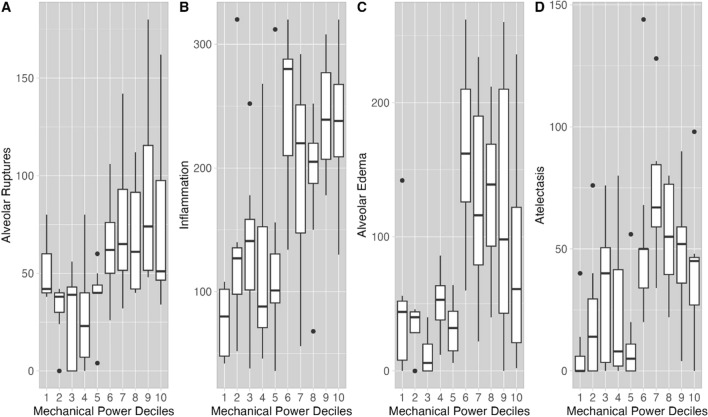
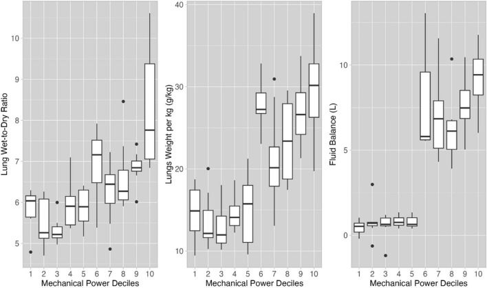
Rationale: Mechanical power (MP) is a summary variable incorporating all causes of ventilator-induced-lung-injury (VILI). We expressed MP as the ratio between observed and normal expected values (MPratio).

Objective: To define a threshold value of MPratio leading to the development of VILI.

Methods: In a population of 82 healthy pigs, a threshold of MPratio for VILI, as assessed by histological variables and confirmed by using unsupervised cluster analysis was 4.5. The population was divided into two groups with MPratio above or below the threshold.

Measurements and main results: We measured physiological variables every six hours. At the end of the experiment, we measured lung weight and wet-to-dry ratio to quantify edema. Histological samples were analyzed for alveolar ruptures, inflammation, alveolar edema, atelectasis. An MPratio threshold of 4.5 was associated with worse injury, lung weight, wet-to-dry ratio and fluid balance (all p < 0.001). After 48 h, in the two MPratio clusters (above or below 4.5), respiratory system elastance, mean pulmonary artery pressure and physiological dead space differed by 32%, 36% and 22%, respectively (all p < 0.001), being worse in the high MPratio group. Also, the changes in driving pressure, lung elastance, pulmonary artery occlusion pressure, central venous pressure differed by 17%, 64%, 8%, 25%, respectively (all p < 0.001).

Limitations: The main limitation of this study is its retrospective design. In addition, the computation for the expected MP in pigs is based on arbitrary criteria. Different values of expected MP may change the absolute value of MP ratio but will not change the concept of the existence of an injury threshold.

Conclusions: The concept of MPratio is a physiological and intuitive way to quantify the risk of ventilator-induced lung injury. Our results suggest that a mechanical power ratio > 4.5 MPratio in healthy lungs subjected to 48 h of mechanical ventilation appears to be a threshold for the development of ventilator-induced lung injury, as indicated by the convergence of histological, physiological, and anatomical alterations. In humans and in lungs that are already injured, this threshold is likely to be different.


Conflict of interest statement

The authors declare that they have no competing interests.

Figures

Fig. 1
Fig. 1
Histological findings. A Severely damaged lung parenchyma with massive alveolar ruptures (H&E, 40×). B Diffuse interalveolar exudation by neutrophils (H&E, 100×). C Lung parenchyma characterized by severe septal blood congestion associated with interalveolar edema (H&E, 40×). D Extensive lung atelectasis involving almost the whole parenchyma in histological section; note also emphysematous area in the left upper corner of the microphotograph (H&E, 40×). E Massive parenchymal blood septal congestion with interalveolar hemorrhages (H&E, 100×). F Extensive blood congestion of septa and vessel of different size (H&E, 40×). G Medium sized artery with edema and focal inflammatory infiltration of the wall (H&E, 40×). H Medium size artery with occlusive recent thrombosis of the lumen; note also marked parenchymal congestion and interalveolar edema (H&E, 40×)
Fig. 2
Fig. 2
Histological damage and mechanical power ratio. Histological score for alveolar ruptures (A), inflammation (B), alveolar edema (C) and atelectasis (D) as a function of mechanical power ratio. This has been expressed in deciles (x-axis) including 8–9 animals each, whose upper limits are the following: decile 1: MPratio 2.40; decile 2: MPratio 2.94; decile 3: MPratio 3.12; decile 4: MPratio 3.54; decile 5: MPratio 4.46; decile 6: MPratio 6.13; decile 7: MPratio 7.28; decile 8: MPratio 10.3; decile 9: MPratio 12.5; decile 10: MPratio 24.6. As shown, upon visual inspection, at MPratio greater than 4.46 (rounded to 4.5, upper limit of the fifth decile), the histological scores markedly increased, suggesting a more severe parenchymal damage (see also Supplement, Table E1)
Fig. 3
Fig. 3
Lung edema and mechanical power ratio. Lung wet-to-dry ratio (A), lung weight (kg−1) (B) and fluid balance (C) as a function of the MPratio deciles. The MPratio limits of each decile are the same as in Fig. 1. Upon visual inspection, at MPratio greater than 4.5, lung wet-to-dry ratio, lung weight and fluid balance also markedly increased
Fig. 4
Fig. 4
Time-course of normalized respiratory system elastance (A), mean pulmonary arterial pressure (B) and physiological dead space (C), in the high (red circles) and low (blue circles) MPratio groups. At the two-way ANOVA analysis: Respiratory system elastance: pGROUP < 0.001, pTIME = 0.039, and pINTERACTION < 0.001. Mean pulmonary artery pressure: pGROUP = 0.003, pTIME < 0.001, and pINTERACTION < 0.001. Physiological dead space: pGROUP = 0.550, pTIME = 0.097, and pINTERACTION = 0.039. See also Table E4 for the complete sets of variables
Fig. 5
Fig. 5
Possible sequences leading to ventilator-induced lung injury in healthy lung. Sequence 1, the intensity of mechanical ventilation is such as to induce structural anatomical modifications, which in turn lead to inflammatory reaction. The physiological variables in theory more associated with these alterations should be the physiological dead space and the inflammatory cytokines, both not specific for lung injury. Sequence 2: the intensity of mechanical ventilation is not such as to induce anatomical damage, but sufficient to interfere with normal hemodynamics. In theory, the first event is the increased intrathoracic pressure followed by all the hemodynamic consequences. The estimate of change of intrathoracic pressure requires the esophageal balloon, while several methods are available to assess the hemodynamic status. Both “direct” and “indirect” lung injury sequences convey in the same final pattern, characterized by water retention, alveolar edema and atelectasis. In the figure we report the common physiological variables which should be more associate with these events

References

    1. Kumar A, Pontoppidan H, Falke KJ, Wilson RS, Laver MB (1973) Pulmonary barotrauma during mechanical ventilation. Crit Care Med 1(4):181–186. 10.1097/00003246-197307000-00001 10.1097/00003246-197307000-00001 - DOI - PubMed
    1. Dreyfuss D, Soler P, Basset G, Saumon G (1988) High inflation pressure pulmonary edema. Respective effects of high airway pressure, high tidal volume, and positive end-expiratory pressure. Am Rev Respir Dis 137(5):1159–1164. 10.1164/ajrccm/137.5.1159 10.1164/ajrccm/137.5.1159 - DOI - PubMed
    1. Tremblay L, Valenza F, Ribeiro SP, Li J, Slutsky AS (1997) Injurious ventilatory strategies increase cytokines and c-fos m-RNA expression in an isolated rat lung model. J Clin Invest 99(5):944–952. 10.1172/JCI119259 10.1172/JCI119259 - DOI - PMC - PubMed
    1. Tobin MJ (2001) Advances in mechanical ventilation. N Engl J Med 344(26):1986–1996. 10.1056/NEJM200106283442606 10.1056/NEJM200106283442606 - DOI - PubMed
    1. Amato MBP et al (2015) Driving pressure and survival in the acute respiratory distress syndrome. N Engl J Med 372(8):747–755. 10.1056/NEJMsa1410639 10.1056/NEJMsa1410639 - DOI - PubMed

LinkOut - more resources