. 2024 Jul 29;15(1):6390.
doi: 10.1038/s41467-024-50807-x.

Harnessing intestinal tryptophan catabolism to relieve atherosclerosis in mice

Affiliations

Harnessing intestinal tryptophan catabolism to relieve atherosclerosis in mice

Mouna Chajadine et al. Nat Commun. .

Abstract

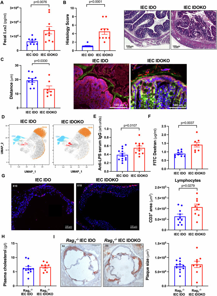
Tryptophan (Trp) is an essential amino acid, whose metabolism is a key gatekeeper of intestinal homeostasis. Yet, its systemic effects, particularly on atherosclerosis, remain unknown. Here we show that high-fat diet (HFD) increases the activity of intestinal indoleamine 2, 3-dioxygenase 1 (IDO), which shifts Trp metabolism from the production of microbiota-derived indole metabolites towards kynurenine production. Under HFD, the specific deletion of IDO in intestinal epithelial cells leads to intestinal inflammation, impaired intestinal barrier, augmented lesional T lymphocytes and atherosclerosis. This is associated with an increase in serotonin production and a decrease in indole metabolites, thus hijacking Trp for the serotonin pathway. Inhibition of intestinal serotonin production or supplementation with indole derivatives alleviates plaque inflammation and atherosclerosis. In summary, we uncover a pivotal role of intestinal IDO in the fine-tuning of Trp metabolism with systemic effects on atherosclerosis, paving the way for new therapeutic strategies to relieve gut-associated inflammatory diseases.


Conflict of interest statement

The authors declare no competing interests.

Figures

Fig. 1
Fig. 1. Induction of IEC IDO by HFD has a protective role in atherosclerosis.
A Tryptophan (Trp), Kynurenine (Kyn) levels, and related Kyn/Trp ratio (%) in the small intestine extracts (n = 5 mice/group), B plasma indole levels in the portal vein, C. 5-hydroxytryptamine (5-HT) in the small intestine extracts of male Ldlr−/− mice fed either normal chow diet (NCD), high-fat diet (HFD), or high-cholesterol diet (HCD) or the combination of both HFD + HCD for 13 weeks (n = 5 mice/group). D Kyn levels in feces of Ldlr−/− mice fed HFD supplemented or not with FOS for 13 weeks (n = 5 mice/group). E, F Kyn/Trp ratio (%) in the small intestine extracts and plasma in male Ldlr−/− IEC IDO KO and littermate control Ldlr−/− IEC IDO mice (n = 5 mice/group), after 8 weeks of HFD + HCD or NCD feeding period. NCD represents the control group without atherosclerosis development. G. Plasma cholesterol, H representative pictures, and quantifications of plaque size in the aortic sinus in male Ldlr−/− IEC IDO KO (n = 12 mice) and littermate control Ldlr−/− IEC IDO (n = 12 mice) fed HFD + HCD for 8 weeks; scale bar 200 µm. I Plasma cholesterol, J representative pictures and quantifications of plaque size in the aortic sinus in female Ldlr−/− IEC IDO KO (n = 9 mice), and littermate controls Ldlr−/− IEC IDO (n = 9 mice) fed HFD + HCD for 13 weeks; scale bar 200 µm. Individual data are presented as scattered dot plots, with the mean and s.e.m. The p values were determined using the Brown-Forsythe one-way ANOVA test followed by Tukey’s multiple comparison test. Source data are provided as a Source Data file.
Fig. 2
Fig. 2. Deficiency of IDO in intestinal epithelial cells (IECs) increases intestinal and plaque inflammation.
A fecal lipocalin 2 (Lcn2) levels scoring in male Ldlr−/− IEC IDO KO (n = 10 mice) and littermate control Ldlr−/− IEC IDO (n = 9 mice). B colon pathohistological scoring in male Ldlr−/− IEC IDO KO and littermate control Ldlr−/− IEC IDO mice (n = 10 mice/group), after 8 weeks of high-fat and high-cholesterol diet (HFD + HCD) feeding period; scale bar 100 µm. C Representative confocal microscopy pictures of microbiota localization: Mucin 2 (green), actin (purple), bacteria (red), and nuclei (blue) and quantifications of mean distances of the closest bacteria (in red) to colonic IEC per condition over three high-powered fields per mouse (IEC IDO n = 10 mice, IEC IDOKO n = 7 mice); scale bar 100 µm. D data were acquired by flow cytometry for Uniform Manifold Approximation and Projection (UMAP) of lymphocytes in the lamina propria of the small intestines. E plasma anti-LPS IgG by ELISA, arbitrary units (arb. units). The results are from male Ldlr−/− IEC IDO KO (n = 11 mice) and littermate control Ldlr−/− IEC IDO mice (n = 13 mice) fed HFD + HCD for 8 weeks. F Detection by ELISA of FITC-dextran in serum of female Ldlr−/− IEC IDO KO (n = 8 mice) and littermate control Ldlr−/− IEC IDO (n = 9 mice) fed HFD + HCD for 13 weeks. G Representative photomicrographs and quantitative analysis of lesional T cells (CD3+ in red) accumulation in the aortic sinus of male Ldlr−/− IEC IDO KO (n = 9 mice) and littermate control Ldlr−/− IEC IDO (n = 10 mice), after 8 weeks of HFD + HCD feeding period; scale bar 100 µm. H plasma cholesterol, I. representative pictures, and quantifications of plaque size in the aortic sinus in male Ldlr−/−Rag1−/− IEC IDO KO (n = 9 mice) and littermate control Ldlr−/−Rag1−/− IEC IDO (n = 11 mice) fed HFD + HCD for 8 weeks; scale bar 200 µm. Individual data are presented as scattered dot plots, with the mean and s.e.m. The p values were determined using the two-tailed Mann-Whitney test. Source data are provided as a Source Data file.
Fig. 3
Fig. 3. Intestinal 5-hydroxytryptamine (5-HT) exhibits pro-inflammatory and pro-atherogenic effects.
A 5-HT levels in small intestine extracts (n = 5 mice/group), B plaque quantification in the aortic sinus and representative images of male Ldlr−/− treated with Tryptophan Hydroxylase 1 (TPH1) inhibitor (LP533401, n = 6 mice) or vehicle (n = 8 mice) and fed HFD + HCD for 8 weeks; scale bar 200 µm. C, D representative images and quantifications of macrophages (MOMA-2+ in red) and lymphocytes (CD3+ in red) accumulation in the aortic sinus (Ldlr−/− Vehicle n = 8, Ldlr−/− LP533401 n = 6); scale bar 100 µm. E representative pictures and quantifications of plaque size in the aortic sinus male Ldlr−/− IEC IDO KO and littermate control Ldlr−/− IEC IDO mice treated with either LP533401 or vehicle and fed HFD + HCD for 8 weeks (IEC IDO Vehicle n = 11 mice, IEC IDO KO Vehicle n = 7 mice, IEC IDO LP533401 n = 11 mice, IEC IDO KO LP533401 n = 9 mice); scale bar 200 µm. F Colon pathohistological scoring (IEC IDO Vehicle n = 5 mice, IEC IDO KO Vehicle n = 10 mice, IEC IDO LP533401 n = 5 mice, IEC IDO KO LP533401 n = 10 mice). G lipocalin-2 (Lcn2) levels in feces (IEC IDO Vehicle n = 5 mice, IEC IDO KO Vehicle n = 7 mice, IEC IDO LP533401 n = 5 mice, IEC IDO KO LP533401 n = 10 mice). Individual data are presented as scattered dot plots, with the mean and s.e.m. The p values were determined using the two-tailed Mann-Whitney test for AD, Kruskal-Wallis, followed by post-Hoc Dunn’s test for F, and one-way ANOVA test followed by Tukey’s multiple comparison test for E and G. Source data are provided as a Source Data file.
Fig. 4
Fig. 4. Trp-dependent microbiota effects impact atherosclerosis.
A Plasma cholesterol, B representative pictures and quantifications of plaques in the aortic sinus of male Ldlr−/− IEC IDO KO and littermate control Ldlr−/− IEC IDO mice treated with antibiotics (ATB) during the 8 weeks of high-fat and high-cholesterol diet (HFD + HCD) feeding period (IEC IDO ATB n = 12 mice, IEC IDO KO ATB n = 11 mice); scale bar 200 µm. C plaque quantification in the aortic sinus of male Ldlr−/− IEC IDO KO and littermate control Ldlr−/− IEC IDO mice either separated by the genotype or mixed (co-housing) in the same cages from the weaning. The mice were fed HFD + HCD for 8 weeks (IEC IDO n = 8 mice, IEC IDO KO n = 7 mice, IEC IDO co-housing n = 8 mice, and IEC IDO KO co-housing n = 8 mice). D colon pathohistological scoring; scale bar 100 µm, E plasma cholesterol levels, F representative pictures and quantifications of plaque size in the aortic sinus; scale bar 200 µm, G representative pictures and quantifications of lymphocytes (CD3+ in red) accumulation within plaques in the aortic sinus of male Ldlr−/−mice treated with 6-Formylindolo(3,2-b)carbazole (Ficz) or vehicle; scale bar 100 µm (n = 8 mice/group) during the 8 weeks of HCD feeding period. Individual data are presented as scattered dot plots, with the mean and s.e.m. The p values were determined using the two-tailed Mann-Whitney test. Source data are provided as a Source Data file.
Fig. 5
Fig. 5. Indole derivatives alleviate inflammation and atherosclerosis.
A indole derivatives (sum of IAA, IPA, and tryptamine) levels supplemented or not in female Ldlr−/− mice fed a high-cholesterol diet (HCD) for 8 weeks (n = 10 mice/group). B data were acquired by flow cytometry for Uniform Manifold Approximation and Projection (UMAP) of leukocytes in the lamina propria of small intestines. Unbiased multi-dimensional analysis by X-shift revealed several different clusters. C plasma cholesterol levels, D representative pictures and plaque size quantification in the aortic sinus; scale bar 200 µm, E representative images and lipid quantification with en-face staining in the thoracic aorta (n = 10 mice/group); scale bar 2 mm, F representative photomicrographs and quantitative analysis of lesional macrophages (MOMA2+ in red) accumulation in the aortic sinus of female Ldlr−/− mice fed HCD for 8 weeks (Ldlr−/− Vehicle n = 9 mice, Ldlr−/− Indole n = 10 mice); scale bar 100 µm. Individual data are presented as scattered dot plots, with the mean and s.e.m. The p values were determined using the two-tailed Mann-Whitney test. Source data are provided as a Source Data file.

References

    1. Cainzos-Achirica, M. et al. Inflammatory Bowel Disease and Atherosclerotic Cardiovascular Disease: JACC Review Topic of the Week. J. Am. Coll. Cardiol.76, 2895–2905 (2020). 10.1016/j.jacc.2020.10.027 - DOI - PubMed
    1. Witkowski, M., Weeks, T. L. & Hazen, S. L. Gut Microbiota and Cardiovascular Disease. Circ. Res127, 553–570 (2020). 10.1161/CIRCRESAHA.120.316242 - DOI - PMC - PubMed
    1. Zelante, T. et al. Tryptophan catabolites from microbiota engage aryl hydrocarbon receptor and balance mucosal reactivity via interleukin-22. Immunity39, 372–385 (2013). 10.1016/j.immuni.2013.08.003 - DOI - PubMed
    1. Gao, J. et al. Impact of the Gut Microbiota on Intestinal Immunity Mediated by Tryptophan Metabolism. Front. Cell. Infect. Microbiol.8, 13 (2018). 10.3389/fcimb.2018.00013 - DOI - PMC - PubMed
    1. Agus, A., Planchais, J. & Sokol, H. Gut Microbiota Regulation of Tryptophan Metabolism in Health and Disease. Cell Host Microbe23, 716–724 (2018). 10.1016/j.chom.2018.05.003 - DOI - PubMed

MeSH terms