. 2024 Jul 30;14(1):17581.
doi: 10.1038/s41598-024-68308-8.

Computer-aided prognosis of tuberculous meningitis combining imaging and non-imaging data

Collaborators, Affiliations

Computer-aided prognosis of tuberculous meningitis combining imaging and non-imaging data

Liane S Canas et al. Sci Rep. .

Abstract

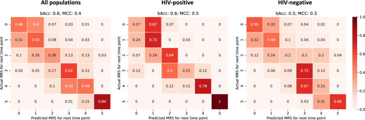
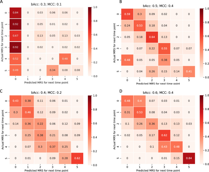
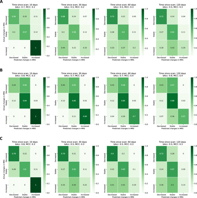
Tuberculous meningitis (TBM) is the most lethal form of tuberculosis. Clinical features, such as coma, can predict death, but they are insufficient for the accurate prognosis of other outcomes, especially when impacted by co-morbidities such as HIV infection. Brain magnetic resonance imaging (MRI) characterises the extent and severity of disease and may enable more accurate prediction of complications and poor outcomes. We analysed clinical and brain MRI data from a prospective longitudinal study of 216 adults with TBM; 73 (34%) were HIV-positive, a factor highly correlated with mortality. We implemented an end-to-end framework to model clinical and imaging features to predict disease progression. Our model used state-of-the-art machine learning models for automatic imaging feature encoding, and time-series models for forecasting, to predict TBM progression. The proposed approach is designed to be robust to missing data via a novel tailored model optimisation framework. Our model achieved a 60% balanced accuracy in predicting the prognosis of TBM patients over the six different classes. HIV status did not alter the performance of the models. Furthermore, our approach identified brain morphological lesions caused by TBM in both HIV and non-HIV-infected, associating lesions to the disease staging with an overall accuracy of 96%. These results suggest that the lesions caused by TBM are analogous in both populations, regardless of the severity of the disease. Lastly, our models correctly identified changes in disease symptomatology and severity in 80% of the cases. Our approach is the first attempt at predicting the prognosis of TBM by combining imaging and clinical data, via a machine learning model. The approach has the potential to accurately predict disease progression and enable timely clinical intervention.

Keywords: DenseNet; Long short-term memory network; MRI imaging; Machine learning; Prognosis; Tuberculous meningitis.


Conflict of interest statement

The authors declare no competing interests.

Figures

Figure 1
Figure 1
Model scheme for individual prognosis prediction. (A:) Feature extractor block, a DenseNet architecture backbone, extracts and encodes the imaging features. The optimisation of this first block is achieved by the contribution of the imaging loss (LI) to the final loss of the model, which assesses the accuracy of the predictions of TBM grade for each MRI scan individually. (B:) Prognosis prediction uses both the clinical (numerical vector) and imaging features (feature maps obtained by the feature extractor block) and uses a bilateral LSTM to predict the mRS scale for the next time-point in the sequence via optimisation of loss of the sequence (LS). The full sequence per individual is assessed. FC: fully connected layer. GT: Ground-true labels. Conv: Convolution layer. Trans: Transition layer. LSTM: Long short-term memory model.
Figure 2
Figure 2
Occlusion sensitivity imaging feature maps for correctly classified patients for coronal, sagittal and axial views (left to right, respectively). (AF): Feature maps for class 0 (TBM grade 1), for HIV-n (AC) and HIV-p (D to F). GM: Feature maps for class 1 (TBM grade 2), for HIV-n (G to I) and HIV-p (K to L). Colormap encodes the relevance of features extracted from the MRI scan, with red encoding highly relevant brain areas for the predicted class and white encoding lower relevance. The occlusion sensitivity maps ranged from 0 to 1, illustrating the probability of each region to highly impact the classification. Black arrows point existent morphological lesions associated to TBM: (A)—tuberculomas and adjacent meningitis, (G)—tuberculomas, (C, L)- vasculitis. Colourmaps were normalised between 0 and 1, with the visualisation threshold set to 0.8.
Figure 3
Figure 3
Confusion matrix of prognosis prediction on the testing set. The confusion matrix is computed considering all time-points of each sequence for the full population, including both HIV-p and HIV-n (left panel), HIV-p (centre) and HIV-n (right panel). bACC: Balanced accuracy. MCC: Mathew correlation score.
Figure 4
Figure 4
Confusion matrix of prognosis prediction on the testing set. The confusion matrix is computed considering all time-points of each sequence for the full population, including both HIV-p and HIV-n. (A): Multivariate prognosis model (MV-PM) using clinical features. (B): Prognosis model using clinical features only. (C): Prognosis model using imaging features only. (D): Proposed prognosis model using both imaging and clinical features. bACC: Balanced accuracy. MCC: Mathew correlation score.
Figure 5
Figure 5
Confusion matrix of disease progression on the testing set given imputed scans. The confusion matrix is computed considering all time-points of each sequence for the full population, including both HIV-p and HIV-n. Increased: the mRS scale increased from the previous timepoint (worsening of patient condition, rise of disease severity). Stable: the mRS scale did not change when compared with the previous timepoint. Decreased: the mRS scale decreased from the previous timepoint (improvement of patient condition). (A): Model trained with clinical data only. The time to scan is measure from the available scan as per the models using imaging, even if not used. (B): Model trained with imaging only. (C): Model trained using imaging and clinical data.

References

    1. Huynh, J. et al. Tuberculous meningitis: Progress and remaining questions. Lancet Neurol21, 450–464 (2022). 10.1016/S1474-4422(21)00435-X - DOI - PubMed
    1. Donovan, J., Thwaites, G. E. & Huynh, J. Tuberculous meningitis: Where to from here?. Curr. Opin. Infect. Dis.33, 259–266. 10.1097/QCO.0000000000000648 (2020). 10.1097/QCO.0000000000000648 - DOI - PMC - PubMed
    1. Berenguer, J. et al. Tuberculous meningitis in patients infected with the human immunodeficiency virus. New England J. Med.326, 668–672 (1992). 10.1056/NEJM199203053261004 - DOI - PubMed
    1. Marais, S. et al. Tuberculous meningitis: A uniform case definition for use in clinical research. Lancet Infect Dis10, 803–812 (2010). 10.1016/S1473-3099(10)70138-9 - DOI - PubMed
    1. Török, M. E. Tuberculous meningitis: Advances in diagnosis and treatment. Br Med. Bull.113, 117–131 (2015). 10.1093/bmb/ldv003 - DOI - PubMed

LinkOut - more resources