Case Reports
. 2024 Jul 30;24(1):867.
doi: 10.1186/s12903-024-04452-x.

Extranodal natural killer/T-cell lymphoma with tonsil involvement: a case report

Affiliations
Case Reports

Extranodal natural killer/T-cell lymphoma with tonsil involvement: a case report

Yang Xiao et al. BMC Oral Health. .

Abstract

Background: Extranodal natural killer/T-cell lymphoma (ENKTL) with tonsil involvement is not common, especially in children.

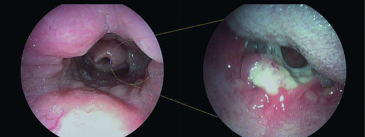
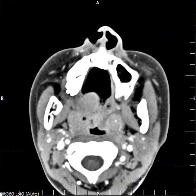
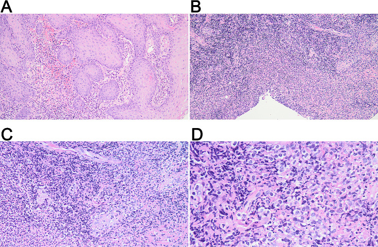
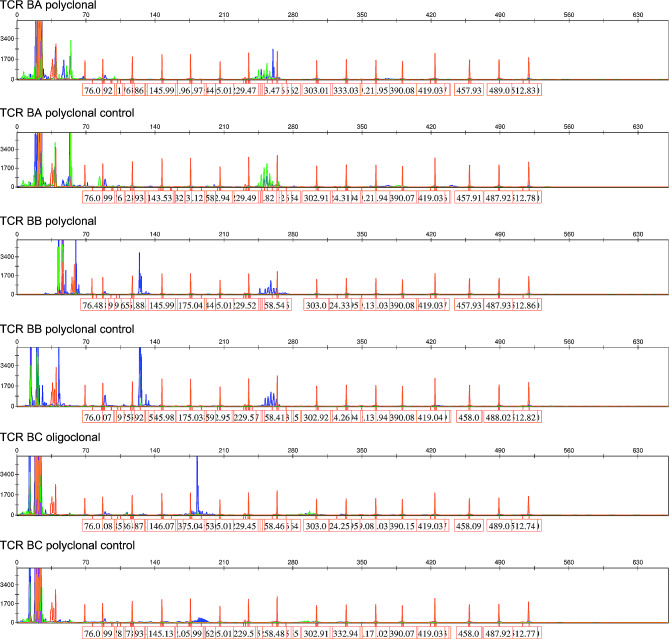
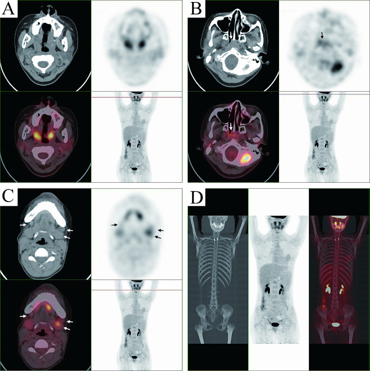
Case presentation: A 13-year-old girl presented with an unexplained sore throat for more than 2 months, together with intermittent fever and suppurative tonsilitis. Nasopharyngoscopy revealed a pharyngeal mass. Enhanced computed tomography (CT) scan showed tonsillar hypertrophy and punctate calcification. Chronic pyogenic granulomatous inflammation with pseudoepithelial squamous epithelial hyperplasia was observed in left tonsil, and pyogenic granulomatous inflammation and a small number of T-lymphoid cells were detected in the right tonsil. The immunohistochemical results showed CD2+, CD3+, CD4+, CD5+, CD8+, granzyme B+, and TIA-1+. The Ki-67 proliferation index was 20%. The case showed T cell receptor gene rearrangement. Finally, the case was diagnosed as ENKTL of stage II with tonsil involvement. The patient received 6 cycles of chemotherapy with SMILE regimen, and showed complete response with no recurrence in the follow-up.

Conclusion: We presented a rare case of ENKTL with tonsil involvement in a child. The patient showed complete response to the SMILE chemotherapy with no recurrence.

Keywords: Case report; ENKTL; Natural killer/T-cell lymphoma; SMILE chemotherapy; Tonsils.


Conflict of interest statement

The authors declare no competing interests.

Figures

Fig. 1
Fig. 1
Nasopharyngoscopy indicated an ulcer lesion in pharyngeal region
Fig. 2
Fig. 2
Contrast enhanced CT showed a slightly swollen uvula, bilateral tonsillar enlargement, and punctate calcification of the left tonsil
Fig. 3
Fig. 3
Representative HE-staining images of tonsil mass. (A) Perilesional pseudoepitheliomatous hyperplasia, under a magnification of 100×; (B) Massive necrosis and vascular proliferation, under a magnification of 100×; (C) Small foci of tumor cells distributed in the necrotic background and invading blood vessels, under a magnification of 200×; (D) Tumor cells have medium, translucent cytoplasm and irregular, distorted nuclei, under a magnification of 400×
Fig. 4
Fig. 4
Immunohistochemical results of tonsil mass. The tumor cells were positive for CD2 (A), CD3 (B), CD4 (C), CD5 (D), CD7 (E), CD8 (F), granzyme B (G), and TIA-1 (H). (I). Ki-67 expression. The images were observed under a magnification of 200×
Fig. 5
Fig. 5
A positive gene scan result of T cell receptor (TCR) gene rearrangement. The clonal rearrangement test utilized PCR-capillary electrophoresis analysis to detect the size distribution of TCR gene amplification. For TCRB-C test, the size range of the PCR amplification product was distributed in the two intervals of 170–210 bp and 285–325 bp. The judgment of monoclonality was based on whether there was a single prominent peak or multiple peaks of similar height within these two ranges. If it was obviously single, it was judged as positive. This TCRB-C obviously had a single blue prominent peak and was judged as positive. For the TCRB-B reaction, the judgment range was 240–285 bp. Within the range, there were multiple blue peaks, and there was no particularly prominent peak. Therefore, it was considered to be negative. NC, negative control
Fig. 6
Fig. 6
Positron emission tomography/computed tomography (PET/CT) indicated infiltration to the throat (A), maxillary (B), and submandibular glands (C). (D) No obvious distant metastasis was detected

References

    1. Reneau JC, Shindiapina P, Braunstein Z, Youssef Y, Ruiz M, Farid S, et al. Extranodal Natural Killer/T-Cell lymphomas: current approaches and future directions. J Clin Med. 2022;11(10):2699. 10.3390/jcm11102699. 10.3390/jcm11102699 - DOI - PMC - PubMed
    1. Locke FL, Ghobadi A, Jacobson CA, Miklos DB, Lekakis LJ, Oluwole OO, et al. Long-term safety and activity of axicabtagene ciloleucel in refractory large B-cell lymphoma (ZUMA-1): a single-arm, multicentre, phase 1–2 trial. Lancet Oncol. 2019;20(1):31–42. 10.1016/s1470-2045(18)30864-7. 10.1016/s1470-2045(18)30864-7 - DOI - PMC - PubMed
    1. Lima M. Aggressive mature natural killer cell neoplasms: from epidemiology to diagnosis. Orphanet J Rare Dis. 2013;8:95. 10.1186/1750-1172-8-95. 10.1186/1750-1172-8-95 - DOI - PMC - PubMed
    1. Sun J, Yang Q, Lu Z, He M, Gao L, Zhu M, et al. Distribution of lymphoid neoplasms in China: analysis of 4,638 cases according to the World Health Organization classification. Am J Clin Pathol. 2012;138(3):429–34. 10.1309/ajcp7yltqpusdq5c. 10.1309/ajcp7yltqpusdq5c - DOI - PubMed
    1. van Doesum JA, Niezink AGH, Huls GA, Beijert M, Diepstra A, van Meerten T. Extranodal Natural Killer/T-cell lymphoma, nasal type: diagnosis and treatment. Hemasphere. 2021;5(2):e523. 10.1097/hs9.0000000000000523. 10.1097/hs9.0000000000000523 - DOI - PMC - PubMed

Publication types

MeSH terms

LinkOut - more resources