

IL-40 is up-regulated in the synovial fluid and cartilage of osteoarthritis patients and contributes to the alteration of chondrocytes phenotype in vitro
- PMID: 39080724
- PMCID: PMC11289996
- DOI: 10.1186/s13075-024-03372-z
IL-40 is up-regulated in the synovial fluid and cartilage of osteoarthritis patients and contributes to the alteration of chondrocytes phenotype in vitro
Abstract
Introduction: IL-40 is a novel cytokine associated with autoimmune connective tissue disorders such as rheumatoid arthritis (RA) or Sjögren syndrome. We have previously shown an accumulation of IL-40 in the RA joint and its expression by immune cells and fibroblasts. Therefore, we aimed to assess the role of IL-40 in association with hyaline cartilage and chondrocyte activity.
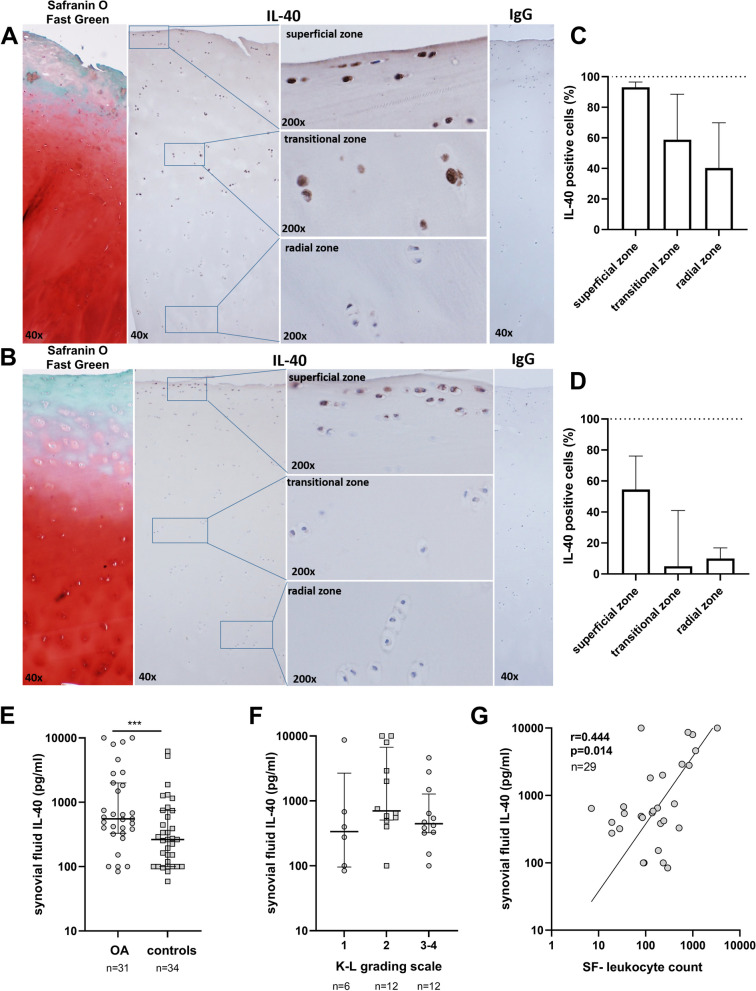
Methods: Immunohistochemistry was employed to detect IL-40 in paired samples of loaded and unloaded regions of osteoarthritis (OA) cartilage (n=5). Synovial fluid IL-40 was analysed by ELISA in OA (n=31) and control individuals after knee injury (n=34). The impact of IL-40 on chondrocytes was tested in vitro.
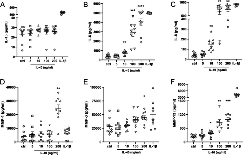
Results: IL-40 was found in chondrocytes of the superficial zone of the OA cartilage, both in loaded and unloaded explants. Additionally, only biopsies from loaded explants showed significant IL-40 positivity in transitional zone chondrocytes. Levels of IL-40 were significantly elevated in the synovial fluid from OA patients compared to controls (p<0.0009) and correlated with synovial fluid leukocyte counts in OA (r=0.444, p=0.014). Chondrocytes exposed to IL-40 dose dependently increased in the secretion of pro-inflammatory cytokines IL-6 (p<0.0001) and IL-8 (p=0.004). Moreover, a dose dependent up-regulation of matrix degrading metalloproteinases MMP-1 (p=0.004), MMP-3 (p=0.031) and MMP-13 (p=0.0002) upon IL-40 treatment was observed in contrast to untreated chondrocytes.
Conclusion: This study is the first to demonstrate the accumulation of IL-40 in OA cartilage and its up-regulation in the synovial fluid of OA patients compared to controls. In addition, extracellular IL-40 appears to play a role in promoting inflammation and cartilage destruction by driving chondrocyte behaviour towards a more aggressive phenotype.
Keywords: Cartilage; Chondrocytes; Interleukin 40; Osteoarthritis.
© 2024. The Author(s).
Conflict of interest statement
The authors declare no competing interests.
Figures
References
-
- Hunter DJ, Bierma-Zeinstra S. Osteoarthritis Lancet. 2019;393:1745–59. - PubMed
MeSH terms
Substances
Grants and funding
- NU21-05-00276/Ministerstvo Zdravotnictví Ceské Republiky
- NU21-05-00276/Ministerstvo Zdravotnictví Ceské Republiky
- NU21-05-00276/Ministerstvo Zdravotnictví Ceské Republiky
- RVO 00023728/Ministerstvo Zdravotnictví Ceské Republiky
- RVO 00023728/Ministerstvo Zdravotnictví Ceské Republiky
- RVO 00023728/Ministerstvo Zdravotnictví Ceské Republiky
- RVO 00023728/Ministerstvo Zdravotnictví Ceské Republiky
- RVO 00023728/Ministerstvo Zdravotnictví Ceské Republiky
- NU21-05-00276/Ministerstvo Zdravotnictví Ceské Republiky
- SVV 260523/Ministry of Education Youth and Sports of the Czech Republic
- SVV 260523/Ministry of Education Youth and Sports of the Czech Republic
- SVV 260523/Ministry of Education Youth and Sports of the Czech Republic
- SVV 260523/Ministry of Education Youth and Sports of the Czech Republic
- SVV 260523/Ministry of Education Youth and Sports of the Czech Republic
- SVV 260523/Ministry of Education Youth and Sports of the Czech Republic
- SVV 260523/Ministry of Education Youth and Sports of the Czech Republic
- SVV 260523/Ministry of Education Youth and Sports of the Czech Republic
- SVV 260523/Ministry of Education Youth and Sports of the Czech Republic
LinkOut - more resources
Full Text Sources
Medical
Molecular Biology Databases
Miscellaneous
