

Early prediction of microvascular invasion (MVI) occurrence in hepatocellular carcinoma (HCC) by 18F-FDG PET/CT and laboratory data
- PMID: 39080787
- PMCID: PMC11290007
- DOI: 10.1186/s40001-024-01973-7
Early prediction of microvascular invasion (MVI) occurrence in hepatocellular carcinoma (HCC) by 18F-FDG PET/CT and laboratory data
Abstract
Background: Hepatocellular carcinoma (HCC) is one of the deadliest malignant tumors in China. Microvascular invasion (MVI) often indicates poor prognosis and metastasis in HCC patients. 18F-FDG PET-CT is a new imaging method commonly used to screen for tumor occurrence and evaluate tumor stage.
Purpose: This study attempted to predict the occurrence of MVI in early-stage HCC through 18F-FDG positron emission tomography (PET)/computed tomography (CT) imaging findings and laboratory data.
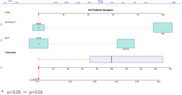
Patients and methods: A total of 113 patients who met the inclusion criteria were divided into two groups based on postoperative pathology: the MVI-positive group and MVI-negative group. We retrospectively analyzed the imaging findings and laboratory data of 113 patients. Imaging findings included tumor size, tumor maximum standard uptake value (SUVmaxT), and normal liver maximum standard uptake value (SUVmaxL). The ratios of SUVmaxT to SUVmaxL (SUVmaxT/L) and an SUVmaxT/L > 2 were defined as active tumor metabolism. The tumor size was indicated by the maximum diameter of the tumor, and a diameter greater than 5 cm was defined as a mass lesion. The laboratory data included the alpha-fetoprotein (AFP) level and the HBeAg level. An AFP concentration > 20 ng/mL was defined as a high AFP level. A HBeAg concentration > 0.03 NCU/mL was defined as HB-positive.
Results: The SUVmaxT/L (p = 0.003), AFP level (p = 0.008) and tumor size (p = 0.015) were significantly different between the two groups. Patients with active tumor metabolism, mass lesions and high AFP levels tended to be MVI positive. Binary logistic regression analysis verified that active tumor metabolism (OR = 4.124, 95% CI, 1.566-10.861; p = 0.004) and high AFP levels (OR = 2.702, 95% CI, 1.214-6.021; p = 0.015) were independent risk factors for MVI. The sensitivity of the combination of these two independent risk factors predicting HCC with MVI was 56.9% (29/51), the specificity was 83.9% (52/62) and the accuracy was 71.7% (81/113).
Conclusion: Active tumor metabolism and high AFP levels can predict the occurrence of MVI in HCC patients.
Keywords: 18F-FDG PET/CT; Hepatocellular carcinoma; Microvascular invasion.
© 2024. The Author(s).
Conflict of interest statement
The authors declare no competing interests.
Figures


Similar articles
-
Preoperative Fluorine 18 Fluorodeoxyglucose Positron Emission Tomography/Computed Tomography for Prediction of Microvascular Invasion in Small Hepatocellular Carcinoma.J Comput Assist Tomogr. 2016 Jul-Aug;40(4):524-30. doi: 10.1097/RCT.0000000000000405. J Comput Assist Tomogr. 2016. PMID: 26966955
-
The value of preoperative 18F-FDG PET metabolic and volumetric parameters in predicting microvascular invasion and postoperative recurrence of hepatocellular carcinoma.Nucl Med Commun. 2022 Jan 1;43(1):100-107. doi: 10.1097/MNM.0000000000001478. Nucl Med Commun. 2022. PMID: 34456318
-
Preoperative prediction of microvascular invasion of hepatocellular carcinoma using 18F-FDG PET/CT: a multicenter retrospective cohort study.Eur J Nucl Med Mol Imaging. 2018 May;45(5):720-726. doi: 10.1007/s00259-017-3880-4. Epub 2017 Nov 22. Eur J Nucl Med Mol Imaging. 2018. PMID: 29167923
-
The importance of a nonsmooth tumor margin and incomplete tumor capsule in predicting HCC microvascular invasion on preoperative imaging examination: a systematic review and meta-analysis.Clin Imaging. 2021 Aug;76:77-82. doi: 10.1016/j.clinimag.2020.11.057. Epub 2020 Dec 3. Clin Imaging. 2021. PMID: 33578134
-
Research progresses of imaging studies on preoperative prediction of microvascular invasion of hepatocellular carcinoma.Clin Hemorheol Microcirc. 2024;88(2):171-180. doi: 10.3233/CH-242286. Clin Hemorheol Microcirc. 2024. PMID: 39031344 Review.
Cited by
-
Evaluating the efficacy of using large language models in preoperative prediction of microvascular invasion in HCC: a multicenter study.Sci Rep. 2025 Jul 29;15(1):27549. doi: 10.1038/s41598-025-08502-4. Sci Rep. 2025. PMID: 40730604 Free PMC article.
-
Diagnostic performance of FDG PET/CT radiomics in predicting microvascular invasion in hepatocellular carcinoma compared to conventional metabolic parameters: a systematic review and meta-analysis.Ann Nucl Med. 2025 Jun 28. doi: 10.1007/s12149-025-02075-y. Online ahead of print. Ann Nucl Med. 2025. PMID: 40581726
References
-
- Lu Y, Chan YT, Wu J, Feng Z, Yuan H, Li Q, Xing T, Xu L, Zhang C, Tan HY, Lee TK, Feng Y, Wang N. CRISPR/Cas9 screens unravel miR-3689a-3p regulating sorafenib resistance in hepatocellular carcinoma via suppressing CCS/SOD1-dependent mitochondrial oxidative stress. Drug Resist Updat Rev Comment Antimicrob Anticancer Chemotherapy. 2023;71: 101015. 10.1016/j.drup.2023.101015.10.1016/j.drup.2023.101015 - DOI - PubMed
-
- Wang S, Zhou L, Ji N, Sun C, Sun L, Sun J, Du Y, Zhang N, Li Y, Liu W, Lu W. Targeting ACYP1-mediated glycolysis reverses lenvatinib resistance and restricts hepatocellular carcinoma progression. Drug Resist Updat Rev Comment Antimicrob Anticancer Chemotherapy. 2023;69: 100976. 10.1016/j.drup.2023.100976.10.1016/j.drup.2023.100976 - DOI - PubMed
-
- Sun B, Ji WD, Wang WC, Chen L, Ma JY, Tang EJ, Lin MB, Zhang XF. Circulating tumor cells participate in the formation of microvascular invasion and impact on clinical outcomes in hepatocellular carcinoma. Front Genet. 2023;14:1265866. 10.3389/fgene.2023.1265866. 10.3389/fgene.2023.1265866 - DOI - PMC - PubMed
MeSH terms
Substances
LinkOut - more resources
Full Text Sources
Medical
Miscellaneous
