. 2024 Sep;60(5):4937-4953.
doi: 10.1111/ejn.16477. Epub 2024 Jul 30.

Cholinergic interneurons in the nucleus accumbens are a site of cellular convergence for corticotropin-releasing factor and estrogen regulation in male and female mice

Affiliations

Cholinergic interneurons in the nucleus accumbens are a site of cellular convergence for corticotropin-releasing factor and estrogen regulation in male and female mice

Kendra L Olson et al. Eur J Neurosci. 2024 Sep.

Abstract

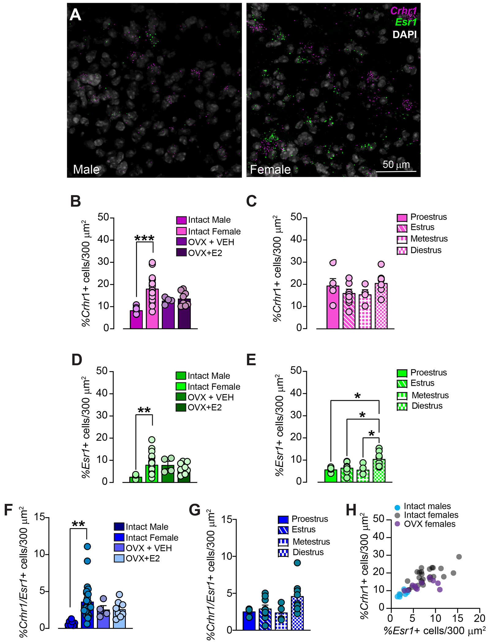
Cholinergic interneurons (ChIs) act as master regulators of striatal output, finely tuning neurotransmission to control motivated behaviours. ChIs are a cellular target of many peptide and hormonal neuromodulators, including corticotropin-releasing factor, opioids, insulin and leptin, which can influence an animal's behaviour by signalling stress, pleasure, pain and nutritional status. However, little is known about how sex hormones via estrogen receptors influence the function of these other neuromodulators. Here, we performed in situ hybridisation on mouse striatal tissue to characterise the effect of sex and sex hormones on choline acetyltransferase (Chat), estrogen receptor alpha (Esr1) and corticotropin-releasing factor type 1 receptor (Crhr1) expression. Although we did not detect sex differences in ChAT protein levels in the dorsal striatum or nucleus accumbens, we found that female mice have more Chat mRNA-expressing neurons than males in both the dorsal striatum and nucleus accumbens. At the population level, we observed a sexually dimorphic distribution of Esr1- and Crhr1-expressing ChIs in the ventral striatum that was negatively correlated in intact females, which was abolished by ovariectomy and not present in males. Only in the NAc did we find a significant population of ChIs that co-express Crhr1 and Esr1 in females and to a lesser extent in males. At the cellular level, Crhr1 and Esr1 transcript levels were negatively correlated only during the estrus phase in females, indicating that changes in sex hormone levels can modulate the interaction between Crhr1 and Esr1 mRNA levels.

Keywords: cholinergic interneuron; corticotropin releasing factor; estrogen receptors; nucleus accumbens.


Figures

Figure 1.
Figure 1.. Region and sex-dependent differences in ChAT-immunoreactivity levels.
A) Schematic diagram of sampling of dorsolateral striatum (DLS), dorsomedial striatum (DMS), nucleus accumbens (NAc) core, and NAc shell across the rostro-caudal axis. B) Example images of ChAT-immunoreactivity (ir; yellow) is seen in neurons (NeuN, magenta) throughout the DLS, DMS, NAc core, and NAc shell. Nuclei indicated with DAPI in grey. Images are maximum projections of 20 μm thick z-stacks. C) Fewer ChAT-ir cells are located in the NAc core compared to the DLS, DMS, or NAc shell (two-way ANOVA, main effect of region, F3,20 = 8.490, p = 0.008), with no differences in number of ChAT-ir cells between males (M, gray, N = 3) and females (F, blue, N = 4; p = 0.1869). D) Percent of ChAT-ir cells that colocalized with the neuronal NeuN marker. ChAT-ir neurons are less dense in the NAc core compared to the DLS, DMS, or NAc shell (two-way ANOVA, main effect of region, F3,20 = 10.66, p = 0.0002) with no effect of sex (p = 0.1427). ###: two-way ANOVA, main effect of region, p < 0.01.
Figure 2.
Figure 2.. Sex differences in Chat mRNA expression.
A) Example images of Chat mRNA visualized in the dorsal striatum (examples from DLS) and NAc (examples from NAc shell) using in situ hybridization alongside DAPI for nucleus visualization. Images are from 5μm thick “single plane” from an example male and female mouse. B) Fewer Chat mRNA+ cells are located in the NAc compared to the DS (two-way ANOVA, main effect of region, F1,40 = 18.11, p = 0.0001, N = 6-26), with females having more Chat+ cells in both the NAc and DS compared to males (two-way ANOVA, main effect of sex, F1,40 = 4.666, p = 0.0368, N = 6-26). Pooled DLS and DMS and pooled NAc core and shell. C) Percent of Chat+ cells co-localized with DAPI. Chat+ cells are less dense in the NAc compared to the DS (two-way ANOVA, main effect of region, F1,40 = 5.839, p = 0.0203), with females having notably denser Chat+ cells in both the NAc and DS compared to males (two-way ANOVA, main effect of sex, F1,40 = 29.26, p < 0.0001, N = 6-26). Pooled DLS and DMS and pooled NAc core and shell. #: Two-way ANOVA main effect of region p < 0.05. ###: Two-way ANOVA main effect of region p < 0.01. $: Two-way ANOVA main effect of sex p < 0.05. $$$$: Two-way ANOVA main effect of sex p < 0.0001.
Figure 3.
Figure 3.. No sex differences in Crhr1+ and Esr1+ mRNA in the dorsal striatum.
A) Example images of in situ hybridization showing Crhr1 (magenta), Esr1 (green), Chat (white), and DAPI (blue) in the male (left) and female (right) dorsal striatum. Examples taken from DLS. B) No change in the density of Crhr1+ cells in the dorsal striatum of males and females (unpaired t-test, t = 0.9380, p = 0.3703, N = 6 males, 6 females). C) The density of Esr1+ cells in the dorsal striatum of males and females is similarly sparse (unpaired t-test, t = 0.5742, p = 0.5785, N = 6 males, 6 females). D) There is minimal overlap in the expression of Crhr1 and Esr1 in the dorsal striatum in male and female mice (unpaired t-test, p = p = 0.5785, N = 6 males, 6 females). E) Percentage of Chat+ neurons (putative cholinergic interneurons, ChIs) co-expressing Crhr1 is similarly high in both male and female mice (unpaired t-test, p = 0.7469). F) Percentage of ChIs co-expressing Esr1 is relatively low in both male and female mice, with female mice showing an increase in inter-animal variability compared to male mice (F test to compare variances, p = 0.0124), but no change in mean percentage of ChIs expressing Esr1 (unpaired t-test, p = 0.1666, N = 6 males, 6 females). G) There is virtually no overlap in the expression of Crhr1 and Esr1 in ChIs in the dorsal striatum in either males or females (unpaired t-test, p = p >0.9999, N = 6 males, 6 females).
Figure 4.
Figure 4.. Sex-dependent differences in Crhr1+ and Esr1+ mRNA in the nucleus accumbens (NAc).
A) In situ hybridization showing Crhr1 mRNA (magenta), Esr1 mRNA (green), and DAPI (white) in male (left) and female (right) NAc. Examples taken from NAc shell. B) The density of Crhr1+ cells is higher in the NAc of females than males (one-way ANOVA, F3,41 = 9.704, p < 0.0001, N = 4–29), with intermediate Crhr1+ cell densities seen in ovariectomized (OVX) females after acute administration of either vehicle (VEH) or 17β-estradiol (E2). C) The density of Crhr1+ cells does not change across phases of the estrous cycle in female mice (one-way ANOVA, F3,21 = 1.463, p = 0.2518, N = 4-9). D) The density of Esr1+ cells is higher in the NAc of females than males, with Esr1+ cellular densities maintained in OVX females treated with either VEH or E2 compared to intact females (one-way ANOVA, F3,43 = 5.073, p = 0.0044, N = 4–29). E) Esr1+ cell density fluctuates over the estrous cycle in intact females, with the highest percentage of Esr1+ cells found during diestrus (one-way ANOVA, F3,21 = 5.560, p = 0.0057, N = 4–9). F) Females have a higher number of cells that co-express Crhr1 and Esr1 in the NAc compared to males, with OVX females showing an intermediate phenotype (one-way ANOVA, F3,41 = 4.490, p = 0.0082, N = 4–29). G) The density of Crhr1+/Esr1+ cells does not change across phases of the estrous cycle in female mice (one-way ANOVA, F3,21 = 2.694, p = 0.0721, N = 4-9) H) There is a positive correlation between the density of Crhr1+ cells and Esr1+ cells in all mice regardless of sex or cycling hormones (intact mice: Pearson’s r = 0.72, p < 0.0001; OVX mice: Pearson’s r = 0.577, p = 0.049). *: Tukey’s post-hoc t-tests, p < 0.05, **: Tukey’s post-hoc t-test, p < 0.01 ***: Tukey’s post-hoc t-test, p < 0.0001
Figure 5.
Figure 5.. Sex-dependent differences in Crhr1+ and Esr1+ co-expression in nucleus accumbens (NAc) cholinergic interneurons (ChIs).
A) Example images of in situ hybridization showing Crhr1 mRNA (magenta), Esr1 mRNA (green), and Chat mRNA (grey, putative ChIs) in male (left) and female (right) NAc. Examples taken from NAc shell. B) Fewer ChIs co-express Crhr1 mRNA in intact females compared to intact males or ovariectomized (OVX) females treated acutely with vehicle (VEH) or 17β-estradiol (E2) (one-way ANOVA, F3,44 = 16.03, p < 0.0001, N = 4–29). C) NAc ChIs express Crhr1 mRNA across the estrous cycle in female mice, with a dip in Crhr1 mRNA expression during diestrus compared to estrus (one-way ANOVA, F3,22 = 3.269, N = 4–9). D) Fewer ChIs co-express Esr1 mRNA in intact males compared to intact females or OVX females treated with either VEH or E2 (one-way ANOVA, F3,44 = 10.47, p < 0.0001, N = 4–29). E) Esr1 mRNA expression in ChIs does not fluctuate across the estrous cycle in intact females (one-way ANOVA, F3,22 = 0.8786, p = 0.4673, N = 4–9). F) Co-expression of Crhr1 with Esr1 mRNA in NAc ChIs is higher in intact females compared to intact males or OVX females treated with either VEH or E2 (one-way ANOVA, F3,44 = 24.72, p < 0.0001, N = 4–29). G) Co-expression of Crhr1 with Esr1 mRNA in NAc ChIs does not fluctuate across the estrous cycle in intact females (one-way ANOVA, F3,22 = 0.4508, p = 0.7193, N = 4–9). H) There is a strong negative correlation between the percentage of ChIs expressing Crhr1 mRNA and the percentage of ChIs expressing Esr1 mRNA in intact female mice (Pearson’s r = −0.63, p < 0.0001), which is not in OVX females (Pearson’s r = 0.12, p > 0.05) or intact males (Pearson’s r = 0.05157, p = 0.9126). I-L) The number of Crhr1 mRNA puncta seen per ChI is not correlated with the number of Esr1 mRNA puncta per ChI during proetrus (I), metestrus (K), or diestrus (L) but is negatively correlated with Esr1 mRNA puncta per ChI during the estrus phase (J) (Pearson’s r = −0.2033, p > 0.0026). *: Tukey’s post-hoc t-test, p < 0.05 **: Tukey’s post-hoc t-test, p < 0.01 ***: Tukey’s post-hoc t-test, p < 0.0001 &: Tukey’s post-hoc t-test, p < 0.1

Update of

Similar articles

Cited by

References

    1. Almey A, Milner TA, Brake WG, 2015. Estrogen receptors in the central nervous system and their implication for dopamine-dependent cognition in females. Hormones and behavior 74, 125–138. - PMC - PubMed
    1. Almey A, Milner TA, Brake WG, 2022. Estrogen receptors observed at extranuclear neuronal sites and in glia in the nucleus accumbens core and shell of the female rat: Evidence for localization to catecholaminergic and GABAergic neurons. J Comp Neurol 530, 2056–2072. - PMC - PubMed
    1. Aoki M, Shimozuru M, Kikusui T, Takeuchi Y, Mori Y, 2010. Sex differences in behavioral and corticosterone responses to mild stressors in ICR mice are altered by ovariectomy in peripubertal period. Zoolog Sci 27, 783–789. - PubMed
    1. Arttamangkul S, Platt EJ, Carroll J, Farrens D, 2021. Functional independence of endogenous mu- and delta-opioid receptors co-expressed in cholinergic interneurons. Elife 10. - PMC - PubMed
    1. Assous M, 2021. Striatal cholinergic transmission. Focus on nicotinic receptors’ influence in striatal circuits. Eur J Neurosci 53, 2421–2442. - PMC - PubMed

LinkOut - more resources