

High-frequency ultrasonography of the scalp: A comparison between androgenetic alopecia and healthy volunteers
- PMID: 39081105
- PMCID: PMC11289427
- DOI: 10.1111/srt.13863
High-frequency ultrasonography of the scalp: A comparison between androgenetic alopecia and healthy volunteers
Abstract
Objective: This study aimed to assess differences in various scalp parameters between patients with androgenetic alopecia (AGA) and healthy volunteers using 22 MHz ultrasound.
Methods: Thirty patients with AGA (AGA group) and 30 healthy volunteers (control group) who visited the Department of Dermatology at the Second Affiliated Hospital of Soochow University from September 2021 to June 2022 were randomly selected. The patients with AGA met the diagnostic criteria outlined in the Chinese Guidelines for the Diagnosis and Treatment of Androgenetic Alopecia. The severity of alopecia was assessed for males between grades 2 and 4 on the Norwood-Hamilton scale, and for females between stages 2 and 3 on the Ludwig scale. No artificial interventions were conducted at the vertex, and all examination conditions remained consistent. Ultrasound examinations at 22 MHz were performed on the scalp at the vertex in both the AGA and control groups. Seven parameters were measured, namely, epidermis + dermis thickness, entire scalp thickness, subcutaneous tissue thickness, average follicle width, average follicle length, follicle count, and the presence of color flow signals in the subcutaneous tissue. The differences in these parameters were then compared.
Results: The AGA group showed reduced thickness of the entire scalp and subcutaneous tissue, narrower average follicle width, shorter average follicle length, lower hair follicle count, and fewer instances of color flow signals in the subcutaneous tissue at the vertex area (p < 0.05).
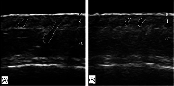
Conclusion: High-frequency (22 MHz) ultrasonography can be employed to visualize the entrance echo, dermis, subcutaneous tissue, and hair follicles of the scalp, thereby providing imaging for the clinical assessment of hair loss.
Keywords: androgenetic alopecia; follicle; scalp; ultrasound.
© 2024 The Author(s). Skin Research and Technology published by John Wiley & Sons Ltd.
Conflict of interest statement
The authors have no conflict of interest to declare.
Figures
References
-
- Alessandrini A, Bruni F, Piraccini BM, Starace M. Common causes of hair loss—clinical manifestations, trichoscopy and therapy. J Eur Acad Dermatol Venereol. 2021;35(3):629‐640. - PubMed
-
- Suchonwanit P, Iamsumang W, Leerunyakul K. Topical finasteride for the treatment of male androgenetic alopecia and female pattern hair loss: a review of the current literature. J Dermatolog Treat. 2022;33(2):643‐648. - PubMed
-
- Panchaprateep R, Tanus A, Tosti A. Clinical, dermoscopic, and histopathologic features of body hair disorders. J Am Acad Dermatol. 2015;72(5):890‐900. - PubMed
-
- Olszewska M, Rudnicka L, Rakowska A, Kowalska‐Oledzka E, Slowinska M. Trichoscopy. Arch Dermatol. 2008;144(8):1007. - PubMed
Publication types
MeSH terms
LinkOut - more resources
Full Text Sources
Miscellaneous
