

Transformation of a Benign Adrenocortical Adenoma to a Metastatic Adrenocortical Carcinoma Is Rare But It Happens
- PMID: 39081696
- PMCID: PMC11288369
- DOI: 10.1210/jcemcr/luae131
Transformation of a Benign Adrenocortical Adenoma to a Metastatic Adrenocortical Carcinoma Is Rare But It Happens
Abstract
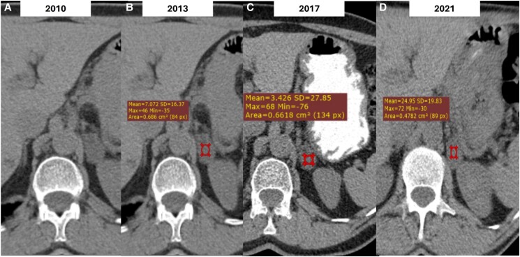
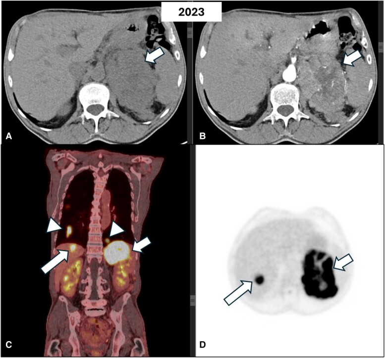
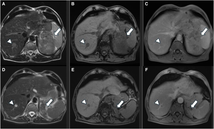
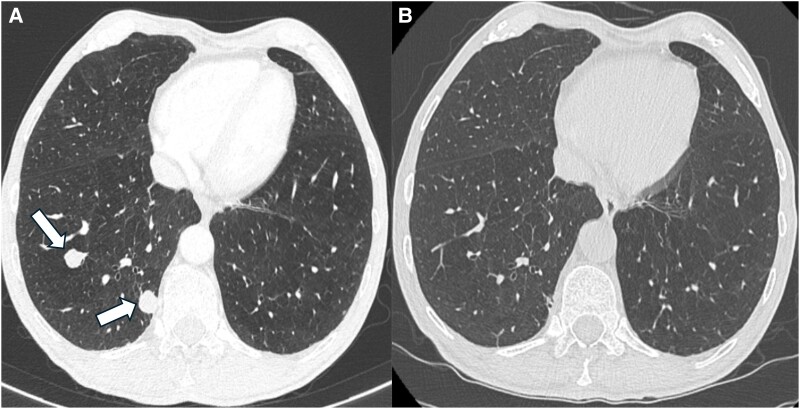
The transformation of an adrenocortical adenoma (ACA) to an adrenocortical carcinoma (ACC) is extremely rare. Current guidelines suggest against further imaging studies and follow-up in patients with nonfunctional adrenal incidentalomas (NFAIs) with benign imaging characteristics. Herein, we present a 64-year-old male patient diagnosed initially with a NFAI of 3 cm in size with imaging characteristics consistent with an ACA. However, 13 years after initial diagnosis, this apparent ACA developed into a high-grade cortisol and androgen-secreting ACC with synchronous metastases. The literature review revealed a further 9 case reports of adrenal incidentalomas initially characterized as ACA that subsequently developed into ACC within a period ranging from 1 to 10 years. The pathogenesis of transformation of an initially denoted ACA to ACC is not fully delineated, although the existing literature focuses on the preexisting or changing genetic background of these lesions, highlighting the need to develop robust prognostic markers to identify patients at risk and individualize the follow-up of these unique cases.
Keywords: adrenal adenoma; adrenal incidentaloma; adrenal incidentalomas; adrenocortical carcinoma; malignant evolution; malignant transformation.
© The Author(s) 2024. Published by Oxford University Press on behalf of the Endocrine Society.
Figures
Similar articles
-
The computed tomography adrenal wash-out analysis properly classifies cortisol secreting adrenocortical adenomas.Endocrine. 2018 Mar;59(3):529-537. doi: 10.1007/s12020-018-1522-7. Epub 2018 Jan 13. Endocrine. 2018. PMID: 29332161
-
Was It an Adrenocortical Adenoma or an Adrenocortical Carcinoma? Limitation of the Weiss Scoring System in Determining the Malignant Potential of Adrenocortical Tumor: Report on Two Cases.Case Rep Endocrinol. 2022 Sep 14;2022:7395050. doi: 10.1155/2022/7395050. eCollection 2022. Case Rep Endocrinol. 2022. PMID: 36160179 Free PMC article.
-
Small adrenal incidentaloma becoming an aggressive adrenocortical carcinoma in a patient carrying a germline APC variant.Endocrine. 2020 Apr;68(1):203-209. doi: 10.1007/s12020-020-02209-4. Epub 2020 Feb 22. Endocrine. 2020. PMID: 32088909
-
Review of Diagnostic Modalities for Adrenal Incidentaloma.J Clin Med. 2023 May 29;12(11):3739. doi: 10.3390/jcm12113739. J Clin Med. 2023. PMID: 37297933 Free PMC article. Review.
-
Giant nonfunctioning adrenal tumors: two case reports and review of the literature.J Med Case Rep. 2018 Nov 10;12(1):335. doi: 10.1186/s13256-018-1876-8. J Med Case Rep. 2018. PMID: 30413177 Free PMC article. Review.
References
-
- Reimondo G, Castellano E, Grosso M, et al. Adrenal incidentalomas are tied to increased risk of diabetes: findings from a prospective study. J Clin Endocrinol Metab. 2020;105(4):973‐981. - PubMed
-
- Mansmann G, Lau J, Balk E, et al. The clinically inapparent adrenal mass: update in diagnosis and management. Endocr Rev. 2004;25(2):309‐340. - PubMed
-
- Fassnacht M, Arlt W, Bancos I, et al. European Society of Endocrinology clinical practice guideline in collaboration with the European network for the study of adrenal tumors. Eur J Endocrinol. 2016;175(2):1‐34. - PubMed
Publication types
LinkOut - more resources
Full Text Sources