. 2024 Sep 9;52(16):9953-9965.
doi: 10.1093/nar/gkae633.

High-throughput determination of RNA tertiary contact thermodynamics by quantitative DMS chemical mapping

Affiliations

High-throughput determination of RNA tertiary contact thermodynamics by quantitative DMS chemical mapping

Bret Lange et al. Nucleic Acids Res. .

Abstract

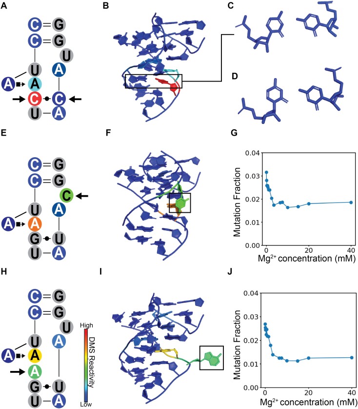
Structured RNAs often contain long-range tertiary contacts that are critical to their function. Despite the importance of tertiary contacts, methods to measure their thermodynamics are low throughput or require specialized instruments. Here, we introduce a new quantitative chemical mapping method (qMaPseq) to measure Mg2+-induced formation of tertiary contact thermodynamics in a high-throughput manner using standard biochemistry equipment. With qMaPseq, we measured the ΔG of 98 unique tetraloop/tetraloop receptor (TL/TLR) variants in a one-pot reaction. These results agree well with measurements from specialized instruments (R2= 0.64). Furthermore, the DMS reactivity of the TL directly correlates to the stability of the contact (R2= 0.68), the first direct evidence that a single DMS reactivity measurement reports on thermodynamics. Combined with structure prediction, DMS reactivity allowed the development of experimentally accurate 3D models of TLR mutants. These results demonstrate that qMaPseq is broadly accessible, high-throughput and directly links DMS reactivity to thermodynamics.


Figures

Graphical Abstract
Graphical Abstract
Figure 1.
Figure 1.
Overview of minimal tetraloop/tetraloop receptor construct. (A) The secondary structure and tertiary structure (PDB: 6DVK) are colored by secondary structure motifs, helices are in black. (B) The secondary and tertiary structures are colored by DMS reactivity, indicating the tetraloop/tetraloop receptor is formed. The 4 adenines that can serve as probes of tertiary contact formation are highlighted in magenta.
Figure 2.
Figure 2.
Buffer effects on the DMS reaction and tertiary contact stability. (A) Using 300 mM sodium cacodylate, compare 40 mM Mg2+ to 0 mM Mg2+ with no notable difference in the TL/TLR mutational fraction. (B) A comparison between 40 mM Mg2+ and 0 mM Mg2+ with 50 mM sodium cacodylate showed notable differences between the mutation fraction in the TL/TLR (highlighted in red), indicating tertiary contact formation. (C) The high correlation between DMS mutation fraction at 50 mM and 300 mM sodium cacodylate at 10 mM Mg2+ indicates that the lower buffer is sufficient to buffer the reaction and gives near identical data. (D) The change in mutation fraction of the GAAA tetraloop between 0 and 40 mM Mg2+ displaying increasing changes as the buffer concentration increases.
Figure 3.
Figure 3.
Nucleotide level conformational information as a function of Mg2+ concentration. (A) 3D structure of the TL/TLR interaction. Colored by residue, red is the three adenines in the tetraloop, green is A4, orange is A5, and blue is A8 of the tetraloop receptor. (A) the 3D structure of the TL/TLR interaction. (B) The secondary structure of the TL/TLR interaction. (C) The mutation fraction of the average of the three adenines of tetraloop in filled circles. The mutation fraction of a TLR-knockout in open circles is missing the TLR (Supplementary Figure S7) and thus cannot form the contact. (D) The mutation fraction of A4 of the TLR in the wild-type (closed circles) and the TL-knockout (open circles). (E) The mutation fraction of A5 of the TLR in the wild-type (closed circles) and the TL-knockout (open circles). (F) The mutation fraction of A8 of the TLR in the wild-type (closed circles) and the TL-knockout (open circles). (G) The proposed three state model of the TLR, A4 (green), A5 (orange), A8 (blue).
Figure 4.
Figure 4.
qMaPseq can measure the destabilization of TL/TLR due to helix lengthening. (A) For a schematic of which helices will be lengthened, see Supplementary Figure S11 for secondary structures of each insertion construct compared to the wild-type. (B) Left is a comparison between the mutation fraction of the wild-type and the 3-bp helix 1 (H1) insertion construct. Middle, a comparison between the mutation fraction of the wild-type and the 3-bp helix 2 (H2) insertion construct. Right, a comparison between the mutation fraction of the wild-type and the 3-bp helix 3 (H3) insertion construct.
Figure 5.
Figure 5.
Comparison between qMaPseq results and known ΔG values for 98 TLR mutations. (A) A schematic of how mutant TLRs were inserted into the miniTTR scaffold. (B) Comparison between Mg2+ titrations of the wild-type and two TLR mutants. (C) Correlation plot between the natural log of [Mg2+]formula image and the ΔG measured by the RNA-MaP experiments from Bonilla et al. for each TLR mutation. (D) Correlation plot between the natural log of average DMS reactivity of the three adenines compared to the ΔG measured by the RNA-MaP experiments from Bonilla et al. for each TLR mutation.
Figure 6.
Figure 6.
DMS reactivity signatures indicate 3D structure features in TLR mutants. (A) DMS reactivity of CCUAAC_CAUGG at 40 mM Mg2+ displaying the unique DMS signature of the C–C mutation highlighted with arrows. (B) FARFAR generated a model of CCUAAC_CAUGG TLR mutant, and the C–C mismatch is boxed. (C) The FARFAR model of the C–C mismatch, which has a unique asymmetrical orientation, with the right C turned to form a hydrogen bond with the N1 position of the left C. (D) The C–C mismatch from the kink-turn motif in the miniTTR scaffold (PDB: 6DVK) has a unique asymmetrical orientation, with the right C turned to form a hydrogen bond with the N1 position of the left C. This asymmetry is likely the cause of its skewed DMS reactivity, where the right C is over 10-fold more reactive than the left C. (E) DMS reactivity of CCUAAG_UACGG with 40 mM Mg2+ with a C9U mutation. (F) In the FARAFAR model of the C9U mutation, this C is flipped out like the wild-type U. (G) The DMS reactivity as a function of Mg2 + where the C levels out to reactivity indicating is consistently solvent accessible. (H) DMS reactivity of CCUAAAG_UAUGG with 40 mM Mg2+ with an A insertion. (I) In the FARAFAR model of the CCUAAAG_UAUGG, the inserted A is flipped out and does not participate in the A–A platform. (J) The DMS reactivity as a function of Mg2+ where the inserted A levels out to reactivity, indicating that it is consistently solvent accessible and has a unique profile that differs from the As in the A–A platform.

References

    1. Bonilla S.L., Denny S.K., Shin J.H., Alvarez-Buylla A., Greenleaf W.J., Herschlag D.. High-throughput dissection of the thermodynamic and conformational properties of a ubiquitous class of RNA tertiary contact motifs. Proc. Natl. Acad. Sci. U.S.A. 2021; 118:e2109085118. - PMC - PubMed
    1. Tian S., Yesselman J.D., Cordero P., Das R.. Primerize: automated primer assembly for transcribing non-coding RNA domains. Nucleic Acids Res. 2015; 43:W522–W526. - PMC - PubMed
    1. Shen W., Le S., Li Y., Hu F.. SeqKit: a cross-platform and ultrafast toolkit for FASTA/Q file manipulation. PLoS One. 2016; 11:e0163962. - PMC - PubMed
    1. Yesselman J.D., Eiler D., Carlson E.D., Gotrik M.R., d’Aquino A.E., Ooms A.N., Kladwang W., Carlson P.D., Shi X.S., Costantino D.A.et al.. Computational design of three-dimensional RNA structure and function. Nat. Nanotechnol. 2019; 14:866–873. - PMC - PubMed
    1. Ban N., Nissen P., Hansen J., Moore P.B., Steitz T.A.. The complete atomic structure of the large ribosomal subunit at 2.4 Å resolution. Science. 2000; 289:905–920. - PubMed

Grants and funding

LinkOut - more resources