. 2024 Aug 20;98(8):e0003524.
doi: 10.1128/jvi.00035-24. Epub 2024 Jul 31.

ORC1 enhances repressive epigenetic modifications on HIV-1 LTR to promote HIV-1 latency

Affiliations

ORC1 enhances repressive epigenetic modifications on HIV-1 LTR to promote HIV-1 latency

Mo Zhou et al. J Virol. .

Abstract

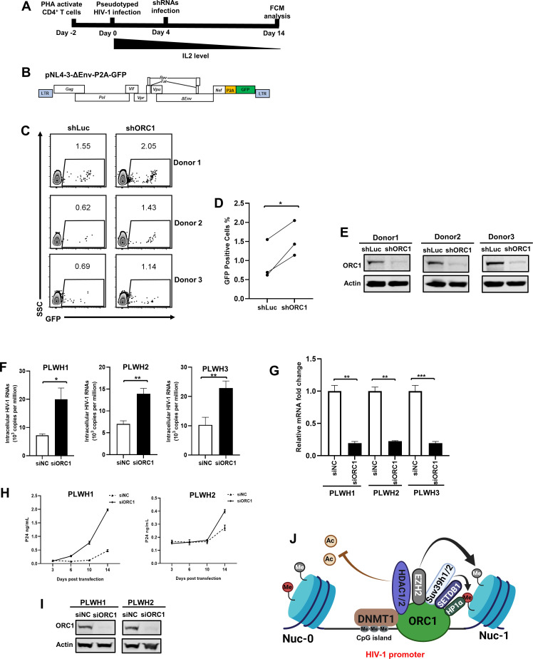
The human immunodeficiency virus type 1 (HIV-1) reservoir consists of latently infected cells which present a major obstacle to achieving a functional cure for HIV-1. The formation and maintenance of HIV-1 latency have been extensively studied, and latency-reversing agents (LRAs) that can reactivate latent HIV-1 by targeting the involved host factors are developed; however, their clinical efficacies remain unsatisfactory. Therefore, it is imperative to identify novel targets for more potential candidates or better combinations for LRAs. In this study, we utilized CRISPR affinity purification in situ of regulatory elements system to screen for host factors associated with the HIV-1 long terminal repeat region that could potentially be involved in HIV-1 latency. We successfully identified that origin recognition complex 1 (ORC1), the largest subunit of the origin recognition complex, contributes to HIV-1 latency in addition to its function in DNA replication initiation. Notably, ORC1 is enriched on the HIV-1 promoter and recruits a series of repressive epigenetic elements, including DNMT1 and HDAC1/2, and histone modifiers, such as H3K9me3 and H3K27me3, thereby facilitating the establishment and maintenance of HIV-1 latency. Moreover, the reactivation of latent HIV-1 through ORC1 depletion has been confirmed across various latency cell models and primary CD4+ T cells from people living with HIV-1. Additionally, we comprehensively validated the properties of liquid-liquid phase separation (LLPS) of ORC1 from multiple perspectives and identified the key regions that promote the formation of LLPS. This property is important for the recruitment of ORC1 to the HIV-1 promoter. Collectively, these findings highlight ORC1 as a potential novel target implicated in HIV-1 latency and position it as a promising candidate for the development of novel LRAs.

Importance: Identifying host factors involved in maintaining human immunodeficiency virus type 1 (HIV-1) latency and understanding their mechanisms prepares the groundwork to discover novel targets for HIV-1 latent infection and provides further options for the selection of latency-reversing agents in the "shock" strategy. In this study, we identified a novel role of the DNA replication factor origin recognition complex 1 (ORC1) in maintaining repressive chromatin structures surrounding the HIV-1 promoter region, thereby contributing to HIV-1 latency. This discovery expands our understanding of the non-replicative functions of the ORC complex and provides a potential therapeutic strategy for HIV-1 cure.

Keywords: HIV-1 latency; LLPS; LTR-associated factors; ORC1; repressive epigenetic modifications.


Conflict of interest statement

The authors declare no conflict of interest.

Figures

Fig 1
Fig 1
ORC1 is identified as a potential host factor associated with the HIV-1 promoter. (A) Schematic of the CAPTURE system employed for screening host factors associated with the HIV-1 promoter. (B) The enrichment of the HIV-1 promoter region in purified chromatin DNA was evaluated by qPCR analysis. The primer set in the reference gene 36B4 served as the control region. Data are presented as means ± SEM in triplicates. P-values were calculated by unpaired Student’s t-test. **P < 0.01. (C) Host factors associated with HIV-1 latency were identified in CAPTURE screen. Conc.(fold enrichment) represents the concentration of proteins in sgLTR group as compared to that in sgNC group. (D–F) J-Lat 10.6 cells were infected with shLuc and shORC1 lentiviruses, respectively. Ninety-six after infection, LRAs including 500 nM SAHA, 1 µM JQ1, and 10 µM Bryostatin-1, were introduced to each group. After 24 hours, the percentage of GFP-positive cells as measured by flow cytometry, as shown in (D). The corresponding statistical results were presented in (E). The knockdown efficiency was validated by Western blot, as shown in (F). Data are presented as means ± SEM in triplicates. P-values were calculated by unpaired Student’s t-test. ***P < 0.001. (G) ChIP assay with antibody against ORC1 was performed in J-Lat 10.6 cells. The qPCR primer positions were presented in the upper panel. The lower panel showed the enrichment of ORC1 on the HIV-1 promoter through qPCR analysis. Data are presented as means ± SEM in triplicates. P-values were calculated by unpaired Student’s t-test. *P < 0.05, **P < 0.01, ***P < 0.001.
Fig 2
Fig 2
ORC1 is associated with repressive epigenetic elements. (A and B) Chromatin accessibility within the HIV-1 proviral genome was assessed in J-Lat 10.6 cells treated with either shLuc or shORC1. Peaks, identified by MACS2, were normalized within each group and visualized using IGV tools. The knockdown efficiency was validated by Western blot, as shown in (B). (C) Hela cells were transfected with Flag-tagged ORC1. The transfection of empty vector was set as control group. Forty-eight hours after transfection, cells from each group were lysed and subsequently immunoprecipitated with anti-Flag beads, accompanied by treatment with nucleases. The enriched proteins were separated by SDS-PAGE and visualized through silver staining. (D) The enriched proteins identified by LC-MS/MS were proceeded to Gene Ontology analysis utilizing PANTHER classification system. (E and F) STRING network analysis was conducted on the enriched proteins with the interaction confidence threshold of 0.7. Clusters involving DNA replication, and chromatin remodeling and organization were identified.
Fig 3
Fig 3
ORC1 regulates the DNA methylation of the HIV-1 promoter by interacting with DNMT1. (A) J-Lat 10.6 cells were lysed and immunoprecipitated with anti-DNMT1 antibody. The endogenous ORC1 enriched by DNMT1 was immunoblotted with anti-ORC1 antibody. (B) HEK293T cells transfected with GFP-tagged ORC1 were treated with AF568-tagged DNMT1 antibody and imaged using super-resolution structured illumination microscopy. (C and D) J-Lat 8.4 cells were infected with shLuc and shORC1 lentiviruses, respectively. Forty-eight hours after infection, cells were treated with 400 nM 5-aza-dC for 1 week. The percentage of GFP-positive cells was measured by flow cytometry, as shown in (C). The corresponding statistical results were presented in (D). Data are presented as means ± SEM in triplicates. P-values were calculated by unpaired Student’s t-test. ***P < 0.001. (E) The upper panel showed the two CpG islands of HIV-1 5′ LTR in J-Lat 8.4 cells. The lower panel presented the CpG methylation profile of HIV-1 5′ LTR in J-Lat 8.4 cells treated as described in (C). In the lower panel, the empty circles represent demethylated sites, while the filled circles represent methylated sites. Statistical significance between groups was determined by Mann-Whitney U test. *P < 0.05, **P < 0.01. (F) ChIP assay with antibody against DNMT1 was performed in shLuc- and shORC1-treated J-Lat 8.4 cells. Data are presented as means ± SEM in triplicates. P-values were calculated by unpaired Student’s t-test. **P < 0.01. (G) The knockdown efficiency of ORC1 in shRNA-infected J-Lat 8.4 cells was validated by Western blot.
Fig 4
Fig 4
HDAC1 and HDAC2 are recruited to the HIV-1 promoter by ORC1. (A) J-Lat 10.6 cells were lysed and immunoprecipitated with anti-HDAC1 antibody. The endogenous ORC1 enriched by HDAC1 was immunoblotted with anti-ORC1 antibody. (B) J-Lat 10.6 cells were lysed and immunoprecipitated with anti-HDAC2 antibody. The endogenous ORC1 enriched by HDAC2 was immunoblotted with anti-ORC1 antibody. (C–G) ChIP assay with antibodies against HDAC1, HDAC2, H3K9ac, and H3K27ac was performed in shLuc- and shORC1-treated J-Lat 10.6 cells. The knockdown efficiency was validated by Western blot, as shown in (G). Data are presented as means ± SEM in triplicates. P-values were calculated by unpaired Student’s t-test. *P < 0.05, **P < 0.01.
Fig 5
Fig 5
ORC1 maintains the repressive histone modifications enriched on the HIV-1 promoter. (A) J-Lat 10.6 cells were lysed and immunoprecipitated with anti-HP1α antibody. The endogenous ORC1 enriched by HP1α was immunoblotted with anti-ORC1 antibody. (B) HEK293T cells transfected with GFP-tagged ORC1 were treated with AF568-tagged antibodies against HP1α, SUV39H1, and H3K9me3, and then imaged using super-resolution structured illumination microscopy (SR-SIM). (C) J-Lat 10.6 cells were lysed and immunoprecipitated with anti-SUV39H1 antibody. The endogenous ORC1 enriched by SUV39H1 was immunoblotted with anti-ORC1 antibody. (D and E) ChIP assay with antibodies against HP1α and H3K9me3 was performed in shLuc- and shORC1-treated J-Lat 10.6 cells. The knockdown efficiency was validated by Western blot, as shown in (E). (F) Flag-tagged ORC1 was transfected in J-Lat 10.6 cells. The endogenous EZH2 enriched by ORC1 was immunoblotted with anti-EZH2 antibody. (G) HEK293T cells transfected with GFP-tagged ORC1 were treated with AF568-tagged antibodies against EZH2 and H3K27me3, and then imaged using SR-SIM. (H and I) ChIP assay with antibody against H3K27me3 was performed in shLuc- and shORC1-treated J-Lat 10.6 cells. The knockdown efficiency was validated by Western blot, as shown in (I).
Fig 6
Fig 6
ORC1 bodies are phase-separated nuclear condensates. (A) Endogenous ORC1 in HEK293T cells was labeled by AF488-tagged antibody and imaged by super-resolution structured illumination microscopy (SR-SIM). (B) The predication of IDRs within ORC1 was performed using PONDR scores. IDRs were subdivided into sub-disordered regions: R1, R2, R3, and R4. The ATPase domain was segmented into four segments: C, D, E, and F. (C) FRAP assay was conducted in living HEK293T cells overexpressing GFP-tagged ORC1. Two ORC1 bodies were selected as regions of interest (ROI). One ROI served as the unbleached negative control, while another ROI was bleached using a high-intensity 488 nm laser. The images were captured every 5 seconds to record the recovery process and changes in fluorescence intensity of ROI. (D) The relative fluorescence intensities of both unbleached and bleached ORC1 bodies in each time point were measured at each time point. The fluorescence intensities at each time point were normalized to the pre-bleaching time point. (E) GFP-tagged ORC1 IDRs were purified in vitro. In the upper panel, 10 µg of proteins was added into Tris-HCl buffer with different NaCl gradients (ranging from 500 to 15.625 mM). In the lower panel, the protein concentration was gradient diluted from 10 µM in Tris-HCl buffer containing 50 mM NaCl. (F) GFP-tagged wild-type ORC1 and LLPS-deficient ORC1 in HEK293T cell were imaged by SR-SIM. (G) Flag-tagged ORC1 and LLPS-deficient ORC1 were transfected in HEK293T-based latency model, respectively. Forty-eight hours after transfection, ChIP assay with anti-Flag antibody was performed. Data are presented as means ± SEM in triplicates. P-values were calculated by unpaired Student’s t-test. **P < 0.01.
Fig 7
Fig 7
ORC1 depletion reactivates latent HIV-1 in cells from people living with HIV-1 (PLWH). (A) The procedure of ORC1 depletion-mediated HIV-1 reactivation in primary CD4+ T cells. (B) The schematic of pseudotyped HIV-1NL4-3-ΔEnv-P2A-GFP backbone. (C–E) The percentage of GFP-positive cells was measured by flow cytometry, as shown in (C). The corresponding statistical results were shown in (D). The knockdown efficiency of ORC1 in shRNA-infected primary CD4+ T cells was validated by Western blot, as shown in (E). Statistical significance between groups was determined by paired Student’s t-test with three replicates per group. *P < 0.05. (F and G) The measurement of HIV-1 cell-associated viral RNA of latently infected CD4+ T cells, which were isolated from PLWH and treated with either siNC or siORC1. The knockdown efficiency of ORC1 in PLWH treated with siRNAs was analyzed by RT-qPCR, as shown in (G). Data are presented as means ± SEM in triplicates. P-values were calculated by unpaired Student’s t-test. *P < 0.05, **P < 0.01, ***P < 0.001. (H–I) The replication-competent HIV-1 was detected by viral outgrowth assay. Primary CD4+ T cells from PLWH were treated with either siNC or siORC1. Irradiated healthy donor peripheral blood mononuclear cells and PHA-activated healthy donor CD4+ T cells were introduced to amplify the reactivated infectious HIV-1. The p24 levels of supernatants at each time point were collected and measured. The knockdown efficiency of ORC1 in PLWH treated with siRNAs was analyzed by Western blot, as shown in (I). (J) The schematic of ORC1-mediated HIV-1 latency.

Similar articles

Cited by

  • HIV-1 latency: From acquaintance to confidant.
    Yang C, Tong L, Xue J. Yang C, et al. J Virus Erad. 2025 May 20;11(2):100597. doi: 10.1016/j.jve.2025.100597. eCollection 2025 Jun. J Virus Erad. 2025. PMID: 40497153 Free PMC article. Review.
  • Grand Challenges on HIV/AIDS in China - The 5th Symposium, Yunnan 2024.
    Chen Z, Xu J, Jin X, Wang J, Huang J, Zhang H, Chen L, Deng K, Cai W, Li L, Wang F, Wu Z, Shang H, Wu H; and the GCC Scientists. Chen Z, et al. Emerg Microbes Infect. 2025 Dec;14(1):2492208. doi: 10.1080/22221751.2025.2492208. Epub 2025 Apr 22. Emerg Microbes Infect. 2025. PMID: 40202047 Free PMC article.

References

    1. Siliciano JD, Kajdas J, Finzi D, Quinn TC, Chadwick K, Margolick JB, Kovacs C, Gange SJ, Siliciano RF. 2003. Long-term follow-up studies confirm the stability of the latent reservoir for HIV-1 in resting CD4+ T cells. Nat Med 9:727–728. doi:10.1038/nm880 - DOI - PubMed
    1. Strain MC, Günthard HF, Havlir DV, Ignacio CC, Smith DM, Leigh-Brown AJ, Macaranas TR, Lam RY, Daly OA, Fischer M, Opravil M, Levine H, Bacheler L, Spina CA, Richman DD, Wong JK. 2003. Heterogeneous clearance rates of long-lived lymphocytes infected with HIV: intrinsic stability predicts lifelong persistence. Proc Natl Acad Sci U S A 100:4819–4824. doi:10.1073/pnas.0736332100 - DOI - PMC - PubMed
    1. Finzi D, Blankson J, Siliciano JD, Margolick JB, Chadwick K, Pierson T, Smith K, Lisziewicz J, Lori F, Flexner C, Quinn TC, Chaisson RE, Rosenberg E, Walker B, Gange S, Gallant J, Siliciano RF. 1999. Latent infection of CD4+ T cells provides a mechanism for lifelong persistence of HIV-1, even in patients on effective combination therapy. Nat Med 5:512–517. doi:10.1038/8394 - DOI - PubMed
    1. Davey RT, Bhat N, Yoder C, Chun TW, Metcalf JA, Dewar R, Natarajan V, Lempicki RA, Adelsberger JW, Miller KD, Kovacs JA, Polis MA, Walker RE, Falloon J, Masur H, Gee D, Baseler M, Dimitrov DS, Fauci AS, Lane HC. 1999. HIV-1 and T cell dynamics after interruption of highly active antiretroviral therapy (HAART) in patients with a history of sustained viral suppression. Proc Natl Acad Sci U S A 96:15109–15114. doi:10.1073/pnas.96.26.15109 - DOI - PMC - PubMed
    1. Bruner KM, Wang Z, Simonetti FR, Bender AM, Kwon KJ, Sengupta S, Fray EJ, Beg SA, Antar AAR, Jenike KM, et al. . 2019. A quantitative approach for measuring the reservoir of latent HIV-1 proviruses. Nature 566:120–125. doi:10.1038/s41586-019-0898-8 - DOI - PMC - PubMed

MeSH terms