Review
. 2024 Sep;68(5):429-442.
doi: 10.1007/s10384-024-01083-1. Epub 2024 Jul 31.

The progress and future of corneal endothelial transplantation

Affiliations
Review

The progress and future of corneal endothelial transplantation

Toshiki Shimizu et al. Jpn J Ophthalmol. 2024 Sep.

Abstract

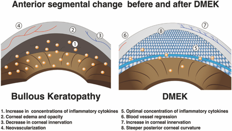
Endothelial transplantation has recently been accepted worldwide, in the long history of corneal transplantation. The introduction of endothelial keratoplasty (Descemet stripping automated endothelial keratoplasty and Descemet membrane endothelial keratoplasty) has enabled us to expand the surgical indications owing to the low incidence of rejection and quick recovery of visual function. New technologies have been developed to ensure stable postoperative outcomes with a shorter learning curve, such as transplantation using cultured human endothelial cells and induced pluripotent stem cells (iPS) or new devices such as artificial endothelium. This review discusses the history and characteristics of corneal transplantation alongside new treatment options that may offer hope for patients with endothelial disease in the future.

Keywords: Corneal endothelial transplantation; Corneal transplantation; DMEK; DSAEK.


Conflict of interest statement

T. Shimizu, None; S. Yamagami, None; T. Hayashi, None.

Figures

Fig. 1
Fig. 1
Illustration of corneal structure. From the superficial layer, the epithelium, Bowman layer, stroma, Descemet membrane, and endothelium are arranged in that order. Dendritic cells are resident in the corneal stroma
Fig. 2
Fig. 2
Anterior segment optical coherence tomography (AS-OCT) photographs of each keratoplasty. a Bullous keratopathy. The corneal stroma is edematous, and the Descemet membrane is visible on the posterior surface of the cornea. b Penetrating keratopathy. The continuity of the cornea is broken. c, d Descemet stripping automated endothelial keratoplasty. The graft is adhered to the posterior surface of the cornea, forming a step. Descemet membrane endothelial keratoplasty. The graft is so thin that it is difficult to recognize it with AS-OCT
Fig. 3
Fig. 3
Illustration of mechanism for antigen presentations. In the direct pathway, donor dendritic cells resident in the corneal graft migrate to the cervical lymph node and present to clusters of differentiation 4 (CD4) cells. In the indirect pathway, a peptide with donor-derived major histocompatibility complex (MHC) antigens is recognized by host antigen-presenting cells (APCs) to T cells
Fig. 4
Fig. 4
Anterior segment photograph after Descemet membrane endothelial keratoplasty. a The transparency of the cornea is remarkably high. b The border between the host endothelium (arrowhead) and the graft (arrow) is visible under high magnification
Fig. 5
Fig. 5
Illustration of the anterior chamber environment before and after DMEK. Before DMEK surgery, the concentration of inflammatory cytokines is high. After DMEK, the anterior chamber environment normalizes owing to the pump mechanism of the corneal endothelium. DMEK Descemet membrane endothelial keratoplasty
Fig. 6
Fig. 6
Illustration of Descemet stripping only. The Descemet membrane is stripped using a reverse Sinskey hook of 4.0 mm in diameter

Similar articles

Cited by

References

    1. Ono T, Ishiyama S, Hayashidera T, Mori Y, Nejima R, Miyata K, et al. Twelve-year follow-up of penetrating keratoplasty. Jpn J Ophthalmol. 2017;61:131–6. - PubMed
    1. Shimazaki J, Amano S, Uno T, Maeda N, Yokoi N. Japan Bullous Keratopathy Study Group. National survey on bullous keratopathy in Japan. Cornea. 2007;26:274–8. - PubMed
    1. Flockerzi E, Maier P, Böhringer D, Reinshagen H, Kruse F, Cursiefen C, et al. Trends in corneal transplantation from 2001 to 2016 in Germany: a report of the DOG-section cornea and its Keratoplasty registry. Am J Ophthalmol. 2018;188:91–8. - PubMed
    1. Armitage WJ, Tullo AB, Larkin DFP. The first successful full-thickness corneal transplant: a commentary on Eduard Zirm’s landmark paper of 1906. Br J Ophthalmol. 2006;90:1222–3. - PMC - PubMed
    1. Tillett CW. Posterior lamellar keratoplasty. Am J Ophthalmol. 1956;41:530–3. - PubMed

MeSH terms

LinkOut - more resources