. 2024 Aug 6;121(32):e2407974121.
doi: 10.1073/pnas.2407974121. Epub 2024 Jul 31.

Targeting the muscarinic M1 receptor with a selective, brain-penetrant antagonist to promote remyelination in multiple sclerosis

Affiliations

Targeting the muscarinic M1 receptor with a selective, brain-penetrant antagonist to promote remyelination in multiple sclerosis

Michael M Poon et al. Proc Natl Acad Sci U S A. .

Erratum in

Expression of concern in

Abstract

Multiple sclerosis (MS) is a chronic and debilitating neurological disease that results in inflammatory demyelination. While endogenous remyelination helps to recover function, this restorative process tends to become less efficient over time. Currently, intense efforts aimed at the mechanisms that promote remyelination are being considered promising therapeutic approaches. The M1 muscarinic acetylcholine receptor (M1R) was previously identified as a negative regulator of oligodendrocyte differentiation and myelination. Here, we validate M1R as a target for remyelination by characterizing expression in human and rodent oligodendroglial cells (including those in human MS tissue) using a highly selective M1R probe. As a breakthrough to conventional methodology, we conjugated a fluorophore to a highly M1R selective peptide (MT7) which targets the M1R in the subnanomolar range. This allows for exceptional detection of M1R protein expression in the human CNS. More importantly, we introduce PIPE-307, a brain-penetrant, small-molecule antagonist with favorable drug-like properties that selectively targets M1R. We evaluate PIPE-307 in a series of in vitro and in vivo studies to characterize potency and selectivity for M1R over M2-5R and confirm the sufficiency of blocking this receptor to promote differentiation and remyelination. Further, PIPE-307 displays significant efficacy in the mouse experimental autoimmune encephalomyelitis model of MS as evaluated by quantifying disability, histology, electron microscopy, and visual evoked potentials. Together, these findings support targeting M1R for remyelination and support further development of PIPE-307 for clinical studies.

Keywords: multiple sclerosis; muscarinic receptors; myelination; oligodendrocyte.


Conflict of interest statement

Competing interests statement:M.M.P., K.I.L., K.J.S., G.C.E., A.R.B., J.R.R., J.M.B., C.S.B., A.C.C., and D.S.L. are current employees of Contineum Therapeutics and hold financial shares of Contineum Therapeutics. Contineum Therapeutics owns patent rights to PIPE-307.

Figures
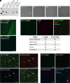
Fig. 1.
Fig. 1.
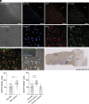
M1R+ OPCs are present in rat and human brain tissue sections. (A) Lysates from CHO-K1 M1R cells or mouse brain were incubated with MT7-CF488A (M1R probe) and separated by native polyacrylamide gel electrophoresis and fluorescent signal detected. Unbound probe migrates at a low molecular weight, below ladder. Specificity was tested using CHO-K1 hM1R membranes, mouse brain homogenate, M1R knockout (M1R KO) and wild-type (M1R WT) homogenates, and CHO-K1 hM1R cell homogenates. Binding was observed in all samples except the M1R KO homogenates. (B) M1R probe (10 nM) binds M1R, but not M2-4R overexpressing CHO-K1 cells. (C) M1R probe competition in mouse hippocampal slice, 100× excess unlabeled MT7 blocks probe binding. (Scale bar: 100 µm.) (D) M1R+/Olig2+/Hoechst+ cells were quantified in various brain regions using human tissue. Top, representative image from a human corpus callosum section showing M1 probe (green), Olig2 (red), and colocalization with Hoechst counterstain (blue). (Scale bar: 25 µm.) Below, summary table of various human brain regions assessed and percentage of Olig2+ and Olig2+/M1R+ cells. All cells quantified were also Hoechst+. (E) Fresh frozen rat cortical sections stained for M1R with the M1R probe (green), then immunostained with the OPC marker PDGFRα, and counterstained with Hoechst (blue). M1R+ OPC (white arrowheads), M1R+ non-OPC (yellow arrowhead). (Scale bar: 50 µm.) (F) Human cerebellar brain section stained with M1R probe (green), then immunostained for the OPC marker NG2 (red), and counterstained with Hoechst (blue). The inset is a magnified image of the OPC highlighted with the white arrowhead. (Scale bar: 25 µm.)
Fig. 2.
Fig. 2.
OPCs expressing M1R are found near MS lesions. (A) Sections of fresh frozen MS donor tissue from the Netherlands Brain Bank were costained with an antibody against NG2 (OPC marker), the MT7-CF488 (M1R probe), and counterstained with Hoechst. Sections were counterstained with the myelin stain Sudan Black to facilitate lesion identification. The dotted yellow line generally outlines the lesion border. Top row are images at 10× magnification (Scale bar: 100 µm.) The region highlighted in the blue box is magnified in the middle row and bottom rows. (B) The blue arrow is approximate region of images highlighted in (A). Characterization of the section was provided by the NBB. The lesion was staged as a 2.3 (active demyelinating lesion containing big rounded foamy HLA+ microglial cells). (C) The percentage of M1R-expressing OPCs (M1R+/NG2+) in non-MS tissue, in MS lesions, as well as lesions classified as “active“, and in lesions classified as “inactive” were quantified. Lesions were characterized by the Netherlands Brain Bank using previously defined criteria.
Fig. 3.
Fig. 3.
Pharmacological profile of PIPE-307 using [3H]-PIPE-307 binding to brain tissue homogenates prepared from adult human cortex and forebrain tissue from adult WT and M1R KO mice. (A and B) Saturation binding was performed using increasing concentrations of [3H]-PIPE-307 in human or mouse brain homogenate. Individual data points represent duplicate samples. (CE) Inhibition binding was performed using 3 nM [3H]-PIPE-307 with increasing concentrations of cold PIPE-307. Individual data points represent the means ± SEM of n = 4 replicates.
Fig. 4.
Fig. 4.
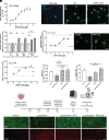
PIPE-307 promotes OPC differentiation and functional remyelination in vitro and in vivo. (A) Rat OPCs were cultured and treated with PIPE-307 for 72 h. Results plotted where T3 induced differentiation was set at 100%. PIPE-307 induces differentiation with an EC50 of 38.6 nM (means ± SEM, n = 18 wells/data point). (B) Images of OPCs treated with 50 ng/mL T3 or 1 µM PIPE-307, MBP (green), Hoechst (blue) (Scale bar: 25 µm.) (C) Rat OPCs were treated with 300 nM PIPE-307 for times indicated then OPCs differentiated until 72 h. White bars indicate vehicle, gray bars PIPE-307 treated, and hatched MT7 treated. PIPE-307 values were normalized to vehicle at respective time point. MT7 was included as a positive control (means ± SD, t test at each time point, n = 5 per point, 2 h: P = 0.65; 8 h: P = 0.003, 24 h: P = 0.01, 8 h: P = 0.0001 for both PIPE-307 and MT7). (D) OPCs differentiated with PIPE-307 wrap axons using a cortical myelination assay. Cultures were immunostained with MBP and myelin segments for each oligodendrocyte measured (myelination index) (means ± SEM, n ≥ 38 cells/data point). (E) Representative image, MBP (green). (Scale bar: 25 µm.) (F) Slice cultures were demyelinated with lysolecithin (Lyso) and then treated with different concentrations of PIPE-307; 300 nM MT7 was used as a positive control. PIPE-307 induces Mbp RNA (measured by qPCR) at an EC50 of 10 nM (means ± SEM, n = 4). (G, H and I) Lysolecithin-treated slice cultures were treated with 300 nM PIPE-307 and then immunostained using antibodies against MBP (myelin) or Caspr and Tuj1 (nodes of Ranvier). The stained MBP area was normalized to Hoechst count. Caspr puncta were normalized to Tuj1 stained area. MT7 was used as a positive control (means ± SEM, ANOVA with Tukey’s n = 4). D, Representative images of slices treated with vehicle alone, or lysolecithin with vehicle, 300 nM MT7, or 1 µM PIPE-307. (Scale bar: 25 µm.)
Fig. 5.
Fig. 5.
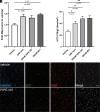
Treatment with PIPE-307 increases Mbp expression and the number of CC1+ oligodendrocytes in human cortical slice cultures. (A) Transcript analysis by qPCR of Mbp mRNA after treatment with 1 µM clemastine (clem), 300 nM PIPE-307, or 300 nM MT7 (means ± SEM, ANOVA with Dunnett’s, n ≥ 6 per group). (B) Quantitation of CC1+/Olig2+ oligodendrocytes following the same treatment as in A (means ± SEM ANOVA with Dunnett’s, n = 4 slices per group). (C) Representative immunohistochemistry of human organotypic cultures using antibodies against Olig2 (red), CC1 (green), and counterstained with Hoechst (blue). (Scale bar: 25 µm.)
Fig. 6.
Fig. 6.
In vivo receptor occupancy profile of PIPE-307. (A) Oral dosing to mouse results in dose-dependent inhibition of total [3H]-PIPE-307 binding in mouse brain (means ± SEM, n ≥ 4/group). The forebrain (FB) was used to determine total binding, a region with high M1 receptor expression. Nonspecific binding was determined using the cerebellum (CB), a region low in M1 receptor expression. (B) Represents the % M1 receptor occupancy of individual subjects plotted as a function dose. PIPE-307 shows a dose-dependent effect with an ED50 = 0.4 mg/kg. (C) Represents the receptor occupancy of individual subjects plotted as a function of plasma and brain PIPE-307 concentrations. The estimated plasma and brain EC50s were similar at 96 nM. The resulting brain to plasma ratio = 1. (D) Time course data following a single oral dose of 3 mg/kg and 30 mg/kg, plotted as % occupancy (as means ± SEM, n ≥ 4/group). 30 mg/kg, PO displays rapid and full occupancy at early time points and falls to ~50% occupancy by 16 h; 3 mg/kg achieves ~50% occupancy for about 10 h postdose. %occupancy = 100 [(treatment specific binding/total baseline specific binding) * 100].
Fig. 7.
Fig. 7.
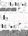
PIPE-307 improves clinical score and induces remyelination in the mouse EAE model. (A), (Left) Daily treatment of PIPE-307 from day 0 decreases the clinical severity in the MOG-EAE model; (Right) cumulative disease index, where scores from day 0 to 22 were summed (means ± SEM; ANOVA with Tukey’s, n = 10 for all groups, ****P < 0.0001, *P < 0.05). (B). PIPE-307 increases the number of myelinated and remyelinated axons in the lumbar spinal cord as defined by a g-ratio > 0.8, but less than 1. (B), left, Bar graph of % total myelinated axons; (B), right, bar graph of % remyelinated axons (means ± SEM, ANOVA with Tukey’s, n = 4 for non-MOG group, n = 10 for all other groups, ***P < 0.001); (B1) scatterplot of g-ratios as a function of axon diameter. (C) Representative electron micrographs of spinal cord tracts from non-MOG-EAE vehicle, MOG-EAE vehicle, and MOG-EAE PIPE-307 (30 mg/kg) treated mice. (D) Treatment with PIPE-307 improves visually evoked potential N1 latency times in the optic nerve (means ± SEM, ANOVA with Dunnett’s post hoc test, n = 16 to 20, ***P < 0.001). Right, Representative traces. (E) PIPE-307 increases the number of total myelinated and remyelinated axons in the optic nerve tract. Left, Bar graph of % total myelinated axons; right, bar graph of remyelinated axons (means ± SEM, ANOVA with Dunnett’s post hoc test, n = 3 for non-MOG, n = 13 to 17, ***P < 0.001); right, scatterplot of g-ratios as a function of axon diameter (means ± SEM, ANOVA, Tukey’s post hoc, n = 3 for non-MOG group, n = 13 to 17 for remaining groups, **P < 0.01, ****P < 0.0001). (F) Representative electron micrographs of optic nerve tracts from non-MOG-EAE vehicle, MOG-EAE vehicle, and MOG-EAE PIPE-307 (30 mg/kg) treated mice.

Comment in

References

    1. Bitsch A., Schuchardt J., Bunkowski S., Kuhlmann T., Bruck W., Acute axonal injury in multiple sclerosis. Correlation with demyelination and inflammation. Brain 123, 1174–1183 (2000), 10.1093/brain/123.6.1174. - DOI - PubMed
    1. Bjartmar C., Wujek J. R., Trapp B. D., Axonal loss in the pathology of MS: Consequences for understanding the progressive phase of the disease. J. Neurol. Sci. 206, 165–171 (2003), 10.1016/s0022-510x(02)00069-2. - DOI - PubMed
    1. Franklin R. J., ffrench-Constant C., Edgar J. M., Smith K. J., Neuroprotection and repair in multiple sclerosis. Nat. Rev. Neurol. 8, 624–634 (2012), 10.1038/nrneurol.2012.200. - DOI - PubMed
    1. Kuhlmann T., et al. , Differentiation block of oligodendroglial progenitor cells as a cause for remyelination failure in chronic multiple sclerosis. Brain 131, 1749–1758 (2008), 10.1093/brain/awn096. - DOI - PubMed
    1. Trapp B. D., Stys P. K., Virtual hypoxia and chronic necrosis of demyelinated axons in multiple sclerosis. Lancet Neurol. 8, 280–291 (2009), 10.1016/S1474-4422(09)70043-2. - DOI - PubMed

MeSH terms

Substances