. 2024 Aug 2;10(31):eadn4682.
doi: 10.1126/sciadv.adn4682. Epub 2024 Jul 31.

ATM and 53BP1 regulate alternative end joining-mediated V(D)J recombination

Affiliations

ATM and 53BP1 regulate alternative end joining-mediated V(D)J recombination

Jinglong Wang et al. Sci Adv. .

Abstract

G0-G1 phase alternative end joining (A-EJ) is a recently defined mutagenic pathway characterized by resected deletion and translocation joints that are predominantly direct and are distinguished from A-EJ in cycling cells that rely much more on microhomology-mediated end joining (MMEJ). Using chemical and genetic approaches, we systematically evaluate potential A-EJ factors and DNA damage response (DDR) genes to support this mechanism by mapping the repair fates of RAG1/2-initiated double-strand breaks in the context of Igκ locus V-J recombination and chromosome translocation. Our findings highlight a polymerase theta-independent Parp1-XRCC1/LigIII axis as central A-EJ components, supported by 53BP1 in the context of an Ataxia-telangiectasia mutated (ATM)-activated DDR. Mechanistically, we demonstrate varied changes in short-range resection, MMEJ, and translocation, imposed by compromising specific DDR activities, which include polymerase alpha, Ataxia-telangiectasia and Rad3-related (ATR), DNA2, and Mre11. This study advances our understanding of DNA damage repair within the 53BP1 regulatory domain and the RAG1/2 postcleavage complex.


Figures
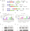
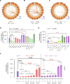
Fig. 1.
Fig. 1.. XRCC1, Parp1, and DDR components support A-EJ of Igκ locus DSBs.
(A) The murine Igκ antigen receptor locus in a germline configuration (GML), each with an associated recombination signal sequence (triangles). STI-571 treatment enables vAbl cells to undergo G0-G1 arrest and initiate V-J recombination. V gene segments are oriented in a deletion (DEL, blue; example: I) or inversion (INV, red; example: II) configuration with respect to the Jκ1 coding end (CE) bait (green arrow). The Jκ1CE can form Vκ coding (CE-CE; I.a or II.a) or hybrid (CE-SE; I.b or II.b) joints with the associated recombination signal end (SE; orange triangles). Recombination efficiency (V-J Eff) was calculated by the sum of DEL and INV V-J joints divided by the total reads. (B) V-J recombination efficiency of Ku70−/− cells with or without inhibitors. (C) V-J recombination efficiency changes of Ku70−/− vAbl cells with added deletions and optionally with ATMi treatment or Ku70 ectopic expression. All experiments were biologically repeated three times, and significance was determined by one-way ANOVA with posttest comparison: *P < 0.05, **P < 0.01, ****P < 0.0001, and ns, no significance; red asterisks indicate significant increases. (D) Western blot of LigIII expression levels in vAbl Ku70−/− (CTR) and Ku70−/− XRCC1−/− (XR1/− #1/2) cells treated by STI-571 for 0, 2, and 4 days (D0, D2, and D4), respectively, where β-actin was used as controls. (E) Same as (D) but for LigI expression levels.
Fig. 2.
Fig. 2.. 53BP1 is essential for G0-G1 A-EJ.
(A) Representative Ku70−/− CE bait junction plots as described in fig. S3B but in the context of 53BP1 single or 53BP1/Exo1 double deletion, with or without ATM inhibition. (B) V-J recombination of the above backgrounds, with or without DNA2, Mre11(ex), and ATM inhibitors, or Ku70 rescue expression. Differences within Ku70−/-53BP1−/− (red bars) and Ku70−/-53BP1−/−Exo1−/− (magenta bars) are evaluated by two-way ANOVA plus posttest comparison: *P < 0.05, ****P < 0.0001; black asterisks indicate significant decreases. All experiments were biologically repeated three times. (A) generated from the Integrative Genomics Viewer (igv.org).
Fig. 3.
Fig. 3.. A-EJ regulators and drivers, but not 53BP1, suppress hyper-resected joints.
(A) Coding and signal prey junctions of Vκ region DSBs from the Jκ1CE bait are aggregated in a resection window of ±100 bp around the RAG1/2 DSB (RSS). Representative plots showing restricted (DNA2i treatment), extended (Polα inhibition; XR1 deletion; Parp1 and ATM inhibition or deletion), or no significant change (53BP1 deletion) are indicated. (B to D) The percentage of junctions that enriched in the window of ±10 bp around the RSS site was used as an indicator for resection, as cyan dashed rectangle in (A). The significance of enrichment changes when combined with indicated inhibitors or gene modification or both was evaluated by one-way ANOVA (B and C) and two-way ANOVA (D) plus posttest comparison; N = 3.
Fig. 4.
Fig. 4.. MMEJ increases with greater declines in A-EJ function.
(A) Jκ1CE bait and Vκ region prey junction structure distributions for the various inhibitors are split into three groups: no detectable pattern change (blue), marginal change (Parpi #1/2, green), and substantial change (Polαi #1/2 and ATMi). The repair profile was shown in the ±10-bp window, the left part indicates overlapping bait/prey microhomology (MH) length, 0 indicates direct (blunt) repair, and the right part indicates the insertion size. (B to D) Same as (A) but for Parp1, 53BP1, and XRCC1 deletion with or without the indicated inhibitors or Ku70 expression, respectively. All experiments were biologically repeated three times.
Fig. 5.
Fig. 5.. ATM and Parp1 maintain recombination fidelity.
(A) An illustration of possible recombination outcomes of post-RAG1/2 cleavage, including coding to coding (CE-CE) (I), signal to signal (SE-SE) (II), and hybrid joins (CE-SE) (III). (B) The Ku70−/− cell CE/SE ratio from Jκ1CE bait is indicated with or without the indicated inhibitors. (C) Same as (B) but with additional XRCC1 (XR1), Parp1, or ATM deletion with or without ATM inhibitor. One-way ANOVA with posttest significance for each comparison is indicated; N = 3. (A) was created with BioRender.com.
Fig. 6.
Fig. 6.. A-EJ and ATM increase distal Vκ recombination.
(A) The Igκ locus is a topologically associated domain (TAD) that can be divided into four subTADs: sTAD1-2, sTAD3, sTAD4, and sTAD5 (53), as shown in the Ku70−/− Jκ1CE bait control. (B) The percentage of the four segments with or without indicated inhibitors as indicated. (C) Same as in (B) but for XRCC1 (XR1), Parp1, and ATM deletions with or without ATM inhibitor. Text highlighted in red and indicated in the graph by red asterisks denotes conditions that extend beyond the ±20% threshold change of the CTR (dashed lines). All experiments represent three biological replications. (A) generated from the Integrative Genomics Viewer (igv.org).
Fig. 7.
Fig. 7.. Compromised DDR affects A-EJ translocations.
(A to C) Representative genome-wide plots of junctions joined with Jκ1CE in Ku70−/− (A), Ku70−/− + ATMi (B), and Ku70−/− 53BP1−/− #1 (C). (D to F) Relative translocation frequencies in Ku70−/− with or without the indicated inhibitors, deletions, or both per 0.5 M (million) sequence read pairs. The significance between Ku70−/− (CTR) and the indicated inhibitors, gene modification, or both was evaluated by one-way ANOVA plus posttest comparison: *P < 0.05, **P < 0.01, ***P < 0.001, ****P < 0.0001; N = 3.
Fig. 8.
Fig. 8.. ATM and 53BP1 regulate A-EJ–mediated V(D)J recombination and chromosome translocation.
Ku70 deficiency eliminates NHEJ and enables a bona fide A-EJ mechanism involving Parp1, XRCC1, and LigIII to complete V(D)J recombination, initiated by the RAG1/2 endonuclease. A-EJ is supported by the ATM-mediated DDR which recruits 53BP1 to facilitate end salvage mechanisms (e.g., distal end tethering) and suppress the formation of translocations (TL). ATM also activates nucleases to promote long-range resection (LR*) and suppress A-EJ when 53BP1 is absent or excluded from DDR recruitment. Thus, inhibiting or deleting ATM (top) increases translocations due to diminished end tethering support and nuclease activation, while deleting 53BP1 (bottom) decreases overall A-EJ capacity by dissociating the V-J tethered repair complex and enabling ATM-activated nucleases to suppress translocations. Created with BioRender.com.

Similar articles

Cited by

References

    1. Zhang Y., Zhang X., Dai H.-Q., Hu H., Alt F. W., The role of chromatin loop extrusion in antibody diversification. Nat. Rev. Immunol. 22, 550–566 (2022). - PMC - PubMed
    1. Schatz D. G., Ji Y., Recombination centres and the orchestration of V(D)J recombination. Nat. Rev. Immunol. 11, 251–263 (2011). - PubMed
    1. Yu W., Lescale C., Babin L., Bedora-Faure M., Lenden-Hasse H., Baron L., Demangel C., Yelamos J., Brunet E., Deriano L., Repair of G1 induced DNA double-strand breaks in S-G2/M by alternative NHEJ. Nat. Commun. 11, 5239 (2020). - PMC - PubMed
    1. Liang Z., Kumar V., Le Bouteiller M., Zurita J., Kenrick J., Lin S. G., Lou J., Hu J., Ye A. Y., Boboila C., Alt F. W., Frock R. L., Ku70 suppresses alternative end joining in G1-arrested progenitor B cells. Proc. Natl. Acad. Sci. U.S.A. 118, e2103630118 (2021). - PMC - PubMed
    1. Liu Q., Palomero L., Moore J., Guix I., Espín R., Aytés A., Mao J.-H., Paulovich A. G., Whiteaker J. R., Ivey R. G., Iliakis G., Luo D., Chalmers A. J., Murnane J., Pujana M. A., Barcellos-Hoff M. H., Loss of TGFβ signaling increases alternative end-joining DNA repair that sensitizes to genotoxic therapies across cancer types. Sci. Transl. Med. 13, eabc4465 (2021). - PMC - PubMed

Publication types

MeSH terms

Substances