An official website of the United States government
The .gov means it’s official.
Federal government websites often end in .gov or .mil. Before
sharing sensitive information, make sure you’re on a federal
government site.
The site is secure.
The https:// ensures that you are connecting to the
official website and that any information you provide is encrypted
and transmitted securely.
1 From the Immunity and Inflammation Theme, Newcastle University Translational and Clinical Research Institute (H.G., S.H.), and the Great North Children's Hospital, Newcastle upon Tyne Hospitals NHS Foundation Trust (P.S.L., E.W., A.M., A.J.C., S.H.), Newcastle upon Tyne, the Department of Clinical Biochemistry and Immunology, Cambridge University Hospital (L.C.-G., S.E., S.D., R.D.), and the National Institute for Health and Care Research (NIHR) Cambridge Biomedical Research Centre (R.D.), Cambridge, the Translational Gastroenterology Unit (N.G., S.T., P.K., H.H.U.), the Kennedy Institute of Rheumatology (S.T.), the NIHR Oxford Biomedical Research Centre (S.T., P.K., H.H.U.), and the Department of Pediatrics (H.H.U.), University of Oxford, Oxford, the Department of Pediatric Gastroenterology, Royal Belfast Hospital for Sick Children (A.S., L.M.), and the Department of Pathology, Royal Victoria Hospital, Belfast Health and Social Care Trust (S.I.), Belfast, and the Department of Pediatric Gastroenterology, Royal Aberdeen Children's Hospital, Aberdeen (S.B.) - all in the United Kingdom; and the Pediatric Gastroenterology Department, Pál Heim National Pediatric Institute, Budapest, Hungary (A.S.).
We discovered high-titer neutralizing autoantibodies against interleukin-10 in a child with infantile-onset inflammatory bowel disease (IBD), a phenocopy of inborn errors of interleukin-10 signaling. After B-cell-depletion therapy and an associated decrease in the anti-interleukin-10 titer, conventional IBD therapy could be withdrawn. A second child with neutralizing anti-interleukin-10 autoantibodies had a milder course of IBD and has been treated without B-cell depletion. We conclude that neutralizing anti-interleukin-10 autoantibodies may be a causative or modifying factor in IBD, with potential implications for therapy. (Funded by the National Institute for Health and Care Research and others.).
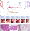
Figure 1. Very early onset inflammatory bowel disease (VEOIBD) in P1, a child with high…
Figure 1. Very early onset inflammatory bowel disease (VEOIBD) in P1, a child with high titer neutralizing autoantibodies to interleukin-10.
(A) Serum anti-IL-10 titer (red, Mean Fluorescence Intensity (MFI) by particle-based assay, 1:100 dilution), IL-10 (purple) and B cell number (blue) over time while receiving therapy as indicated in (B); EN, exclusive enteral nutrition; PN, parenteral nutrition; IVIG, intravenous immunoglobulin (high dose/replacement dose indicated by height of bar). (C) Clinical timeline in which the severity of GI symptoms is indicated by the height of the pink shading. Asterisks show the timing of endoscopic examinations. Triangles at 52 (blue) and 84 (green) months represent brief episodes of oedema and altered fecal calprotectin, respectively, as described in the text. (D) Sequential colonoscopic appearances showing improvement in ulceration (arrows) over time, left to right. (E) Representative images of colonic histopathology at indicated time-points, stained with hematoxylin and eosin. Left: mild distortion of glandular architecture associated with increased cellularity of the lamina propria and admixed acute and chronic inflammation at 20-26 m of age. Right: progressive resolution of inflammatory changes & restoration of normal architecture.
Figure 2. Identification and functional characterization of…
Figure 2. Identification and functional characterization of autoantibodies to interleukin-10 in 2 children with VEOIBD.
A: IL-10-mediated downregulation of LPS induced TNF alpha in Whole Blood (WB) and PBMC. Whole blood and PBMCs of healthy controls (WB n=50, PBMC n = 3) and patients (P1, P2) were activated with lipopolysaccharide (LPS) alone or in the presence of IL-10. Addition of LPS induces strong up-regulation of TNF alpha production when compared to non-stimulated samples (ns). Control WB and PBMC show significant downregulation of LPS-induced TNFα production when co-stimulated with IL-10, while there is no downregulation in WB of P1 and P2. Patient responses are restored when stimulating PBMC in the absence of autologous serum. Patient P1’s WB was also tested after treatment, when anti-IL-10 had declined (“Post”), showing a normal response similar to controls. Mean values with standard deviations (SD) are shown. IL10-mediated downregulation of WB controls was analysed by Mann-Whitney test. B: Titration of anti-IL-10 IgG. Sera of P1 and P2 were tested by particle-based flow cytometry for the presence of auto-antibodies to IL-10. Titrations for anti-IL-10 IgG were performed in 1/5 dilution steps starting at a dilution of 1/20. Titers at initial presentation and post treatment are shown for P1 and P2 (mean values +/- SD from duplicate testing). 20 healthy adult controls (mean +/- SD) are as well shown at dilutions 1/20 and 1/100. Results from an additional cohort of 50 healthy pediatric control sera at a dilution of 1/100 are shown on the right side. C: IL-10 reporter assay (HEK-blue™ IL-10 cells). An IL-10 responder line was activated with IL-10 either alone (4 ng/ml, n=5, white bars/circles) or in the presence of control serum (gray bars/circles, n=10) or patient serum P1 (red bars/triangles, 1:10, n=3) or P2 (red bars/circles, 1:10, n=3). Sera of P1 and P2 significantly inhibit IL-10-induced SEAP induction (Mann-Whitney, p < 0.0002) as determined by colorimetric measurement at OD620. D: IL-10-induced STAT3 activation in a dual luciferase reporter assay. Effect of patient or control serum on IL-10-mediated STAT3 transcriptional activity. Sera were tested for their ability to interfere with IL-10 induced STAT3 activation of a reporter-transfected cell line, measured by luciferase induction (Mann-Whitney, p < 0.0055). Activation was performed either with IL-10 alone (200 pg/ml), or in the presence of control serum (gray bar/circles, n=10) or patient serum of P1 (red bar/triangle, 1:10) or P2 (red bars/circles, 1:10). E: IL-10 cross-inhibition assay. PBMC of patient P2 and a healthy control (C) were activated by LPS alone or LPS + IL-10, in the presence of either control serum or P2 serum. Comparison is made with responses in whole blood (WB). Bars indicated mean values of 2 replicates from 1 experiment, symbols show individual values. P2 serum blocks IL-10-mediated downregulation of TNF alpha by control and patient cells.
Glocker E-O, Kotlarz D, Boztug K, et al. Inflammatory bowel disease and mutations affecting the interleukin-10 receptor. New England Journal of Medicine. 2009;361(21):2033–2045.
-
PMC
-
PubMed
Glocker E-O, Frede N, Perro M, et al. Infant colitis—it’s in the genes. The Lancet. 2010;376(9748):1272.
-
PubMed
Sazonovs A, Stevens CR, Venkataraman GR, et al. Large-scale sequencing identifies multiple genes and rare variants associated with Crohn’s disease susceptibility. Nature genetics. 2022;54(9):1275–1283.
-
PMC
-
PubMed
Zhao JH, Stacey D, Eriksson N, et al. Genetics of circulating inflammatory proteins identifies drivers of immune-mediated disease risk and therapeutic targets. Nat Immunol. 2023;24(9):1540–1551. doi: 10.1038/s41590-023-01588-w.
-
DOI
-
PMC
-
PubMed
Tangye SG, Al-Herz W, Bousfiha A, et al. Human inborn errors of immunity: 2022 update on the classification from the international union of immunological societies expert committee. Journal of clinical immunology. 2022;42(7):1473–1507.
-
PMC
-
PubMed