. 2024 Dec;167(7):1399-1414.e2.
doi: 10.1053/j.gastro.2024.07.025. Epub 2024 Jul 30.

Mucosal Single-Cell Profiling of Crohn's-Like Disease of the Pouch Reveals Unique Pathogenesis and Therapeutic Targets

Affiliations

Mucosal Single-Cell Profiling of Crohn's-Like Disease of the Pouch Reveals Unique Pathogenesis and Therapeutic Targets

Siyan Cao et al. Gastroenterology. 2024 Dec.

Abstract

Background & aims: The pathophysiology of Crohn's-like disease of the pouch (CDP) in patients with a history of ulcerative colitis (UC) is unknown. We examined mucosal cells from patients with and without CDP using single-cell analyses.

Methods: Endoscopic samples were collected from pouch body and prepouch ileum (pouch/ileum) of 50 patients with an ileal pouch-anal anastomosis. Single-cell RNA sequencing was performed on pouch/ileal tissues of patients with normal pouch/ileum and CDP. Mass cytometry was performed on mucosal immune cells from patients with UC with normal pouch/ileum, CDP, pouchitis, and those with familial adenomatous polyposis after pouch formation. Findings were independently validated using immunohistochemistry.

Results: The cell populations/states in the pouch body differed from those in the prepouch ileum, likely secondary to increased microbial burden. Compared with the familial adenomatous polyposis pouch, the UC pouch was enriched in colitogenic immune cells even without inflammation. CDP was characterized by increases in T helper 17 cells, inflammatory fibroblasts, inflammatory monocytes, TREM1+ monocytes, clonal expansion of effector T cells, and overexpression of T helper 17 cells-inducing cytokine genes such as IL23, IL1B, and IL6 by mononuclear phagocytes. Ligand-receptor analysis further revealed a stromal-mononuclear phagocytes-lymphocyte circuit in CDP. Integrated analysis showed that up-regulated immune mediators in CDP were similar to those in CD and pouchitis, but not UC. Additionally, CDP pouch/ileum exhibited heightened endoplasmic reticulum stress across all major cell compartments.

Conclusions: CDP likely represents a distinct entity of inflammatory bowel disease with heightened endoplasmic reticulum stress in both immune and nonimmune cells, which may become a novel diagnostic biomarker and therapeutic target for CDP.

Keywords: CyTOF; IBD; IPAA; Pouch; scRNA-seq.


Conflict of interest statement

Conflicts of interest

The authors disclose no conflicts.

Figures
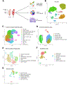
Figure 1.
Figure 1.
Mucosal single-cell landscape of pouch body and prepouch ileal biopsy specimens from patients with UC. (A) Workflow of sample collection from normal pouch/ileum (UC) and CDP for scRNA-seq with B cell receptor (BCR)/TCR-seq, CyTOF, and IHC. FAP or pouchitis samples were not included in this panel for simplicity. (B) Uniform Manifold Approximation and Projection (UMAP) visualization of all cells in the pouch body and prepouch ileum from patients with a normal pouch/ileum (UC) and CDP. (CG) UMAP visualization of detailed cell types within each of the 5 major compartments from patients with a normal pouch/ileum (UC) and CDP. cDC, conventional dendritic cells; GC, germinal center; Tcm, central memory T cells; Tem, effector memory T cells.
Figure 2.
Figure 2.
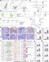
Skewed mucosal cell composition and pathways in normal pouch body from UC patients. (A) Subtype composition of MNPs, T cells and innate lymphoid cells (s), epithelial cells, and stromal cells from normal pouch body (UC) vs normal prepouch ileum (UC) by scRNA-seq (n = 5). (B) Pathway analysis of antimicrobial immune response in normal pouch body (UC) relative to prepouch ileum (UC) using Single Cell Pathway Analysis (SCPA). (C) Ligand-receptor analysis of normal pouch body (UC) relative to normal prepouch ileum (UC) using MultiNicheNet. (D) Pathway analysis of epithelial cell metabolism in normal pouch body (UC) relative to normal prepouch ileum (UC) using SCPA. (E) IHC of proteins encoded by altered epithelial cell genes in normal pouch body (UC) vs normal pre-pouch ileum (UC) (n = 6). Scale bars: 50 μm. Immune cell frequencies of (F) normal pouch body and (G) normal prepouch ileum from patients with UC or FAP after IPAA by CyTOF (n = 5). cDC, conventional dendritic cells; NL, normal; Mac, macrophage. Error bars indicate standard error of the mean. P values were calculated using the Mann-Whitney U test. *P < .05, **P < .01.
Figure 3.
Figure 3.
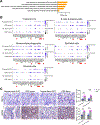
Analysis by scRNA-seq and CyTOF uncovers rewired immune and nonimmune landscapes in CDP. (A) Uniform Manifold Approximation and Projection (UMAP) visualization of all cells from CDP and normal pouch/ileum (UC) (n = 5 or 6). ILC, innate lymphocyte cell. Frequencies of each individual cell populations in CDP vs normal pouch/ileum (UC) in the (B) pouch body and (C) prepouch ileum by scRNA-seq (n = 5 or 6). Cell-type frequencies of all cells in a sample are shown. NL, normal. (D) Ig isotypes of plasma cells from CDP and normal pouch/ileum (UC) by B cell receptor-seq (n = 5 or 6). (E) Immune cell populations of normal pouch/ileum (UC), CDP, and pouchitis analyzed by CyTOF (n = 9 or 10). Error bars indicate standard error of the mean. P values were calculated using the Mann-Whitney U test. *P < .05, **P < 0.01.
Figure 4.
Figure 4.
CDP exhibits expanded TCR clonotypes, activated Th17 signaling, and decreased T cell exhaustion. (A) T cell clonal expansion in normal pouch/ileum (UC) and CDP by TCR-seq (n = 5 or 6). (B) Clonal expansion of T cell subpopulations by TCR-seq. (C) Uniform Manifold Approximation and Projections (UMAPs) of T cell clusters (left) as well as TCR signaling scores in CDP ileum and CDP pouch by TCR-seq. (D) Expression of exhaustion markers in CD8 T cells from the pouch body and prepouch ileum of normal pouch/ileum (UC) and CDP by scRNA-seq.
Figure 5.
Figure 5.
Upregulation of inflammatory mediators in MNPs and fibroblasts in CDP by scRNA-seq. (A) MNPs from CDP tissues enriched genes encoding proinflammatory and proremodeling mediators compared with normal (NL) pouch/ileum (UC) by scRNA-seq (n = 5 or 6). (B) Enrichment of inflammatory monocytes in CDP compared with normal pouch/ileum (UC) (n = 5 or 6). (C) Cross-validation of the cluster of inflammatory monocytes using markers identified by Smillie et al. (D) Elevations in Th1/Th17-inducing cytokines by MNPs in CDP tissues. (E) Enrichment of CD14 and TREM1 in monocytes from CDP compared with normal pouch/ileum (UC) by scRNA-seq. (F) Uniform Manifold Approximation and Projection (UMAP) visualization shows elevated inflammatory fibroblasts in CDP tissues. (G) Elevated expression of chemokine genes CCL13, CCL8, CXCL6, and CCL2 in inflammatory fibroblasts relative to other stromal cells in the pouch body and prepouch ileum of CDP. (H) IHC of inflammatory mediators IL-1β and CCL2 in CDP and normal pouch/ileal (UC) tissues (n = 6). Scale bars: 50 µm. (I) Ligand-receptor interactions in CDP and normal pouch/ileum (UC) using MultiNicheNet. (J and K) T cells isolated from CDP and normal pouch/ileal (UC) samples were stimulated with IL23 and IL-1β ex vivo. Th17 cells and intracellular cytokines were identified by flow cytometry. Error bars indicate standard error of the mean. P values were calculated using the Mann-Whitney U test. *P < .05, **P < .01.
Figure 6.
Figure 6.
Integrated analyses of mucosal cells in UC, CD, pouchitis, and CDP. (A) Frequencies of cell subsets in uninflamed pouch and pouchitis, as well as normal pouch/ileum (UC) (NL) and CDP by integrated analysis. (B) Frequencies of cell subsets in healthy controls, UC (uninflamed and inflamed), CD (uninflamed and inflamed), and normal pouch/ileum (UC) (NL) and CDP. (C) Monocyte diffusion plot in pouch body of CDP vs pouchitis by integrated pseudotime analysis. DC, diffusion component. (D) Monocyte distribution along pseudotime in pouch body of CDP vs pouchitis. Expression of proinflammatory genes in (E and F) CDP, pouchitis, UC, and CD in MNPs and (G) stromal cells by integrated analysis. Tc17 cells, IL17+ CD8 T cells. Error bars indicate standard error of the mean. *P < .05.
Figure 7.
Figure 7.
UPR in all major cell compartments is a unique feature of CDP. (A) Pathway analysis of ER stress and the UPR pathways in CDP vs normal pouch/ileum (UC) using Single Cell Pathway Analysis (SCPA). (B) UPR gene expression in major cell compartments in CDP tissues and normalpouch/ileum (UC) (NL). (C) IHC of XBP1 and HSPA5 in pouch/ileal samples of an independent patient cohort of normal pouch/ileum (UC) and CDP. Arrowheads point to nuclear staining of XBP1. Scale bars: 50 µm. Error bars indicate standard error of the mean. P values were calculated using the Mann-Whitney U test. **P < .01, ***P < .001.

References

    1. Akiyama S, Rai V, Rubin DT. Pouchitis in inflammatory bowel disease: a review of diagnosis, prognosis, and treatment. Intest Res 2021;19:1–11. - PMC - PubMed
    1. Barnes EL, Kochar B, Jessup HR, et al. The incidence and definition of Crohn’s disease of the pouch: a systematic review and meta-analysis. Inflamm Bowel Dis 2019;25:1474–1480. - PMC - PubMed
    1. Shen B, Kochhar GS, Kariv R, et al. Diagnosis and classification of ileal pouch disorders: consensus guidelines from the International Ileal Pouch Consortium. Lancet Gastroenterol Hepatol 2021;6:826–849. - PubMed
    1. Shen B, Kochhar GS, Rubin DT, et al. Treatment of pouchitis, Crohn’s disease, cuffitis, and other inflammatory disorders of the pouch: consensus guidelines from the International Ileal Pouch Consortium. Lancet Gastroenterol Hepatol 2022;7:69–95. - PubMed
    1. Cao SY, Cella M, Jaeger N, et al. Mass cytometry analysis of Crohn’s disease-like phenotype of the ileoanal pouch following ileal pouch-anal anastomosis for ulcerative colitis. Gastroenterology 2022;162:S58–S59.

MeSH terms