Review
. 2024 Jul;31(4):317-325.
doi: 10.1053/j.akdh.2023.12.007.

An Update on Viral Infection-Associated Collapsing Glomerulopathy

Affiliations
Review

An Update on Viral Infection-Associated Collapsing Glomerulopathy

Molly Fisher et al. Adv Kidney Dis Health. 2024 Jul.

Abstract

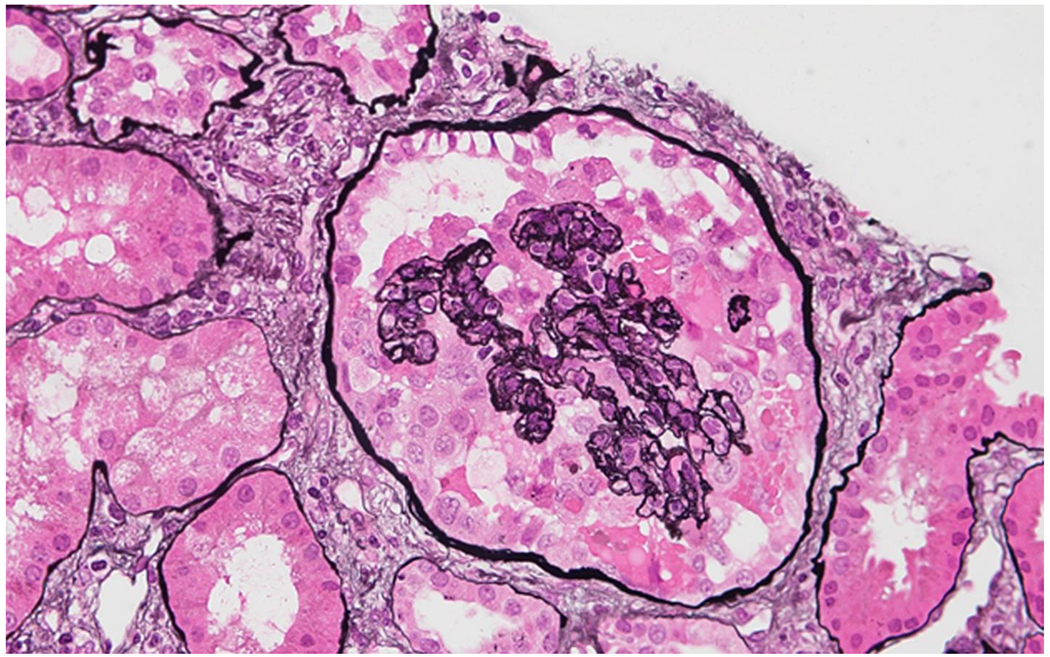
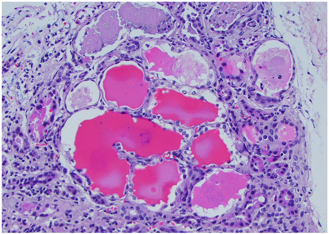
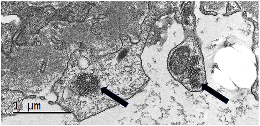
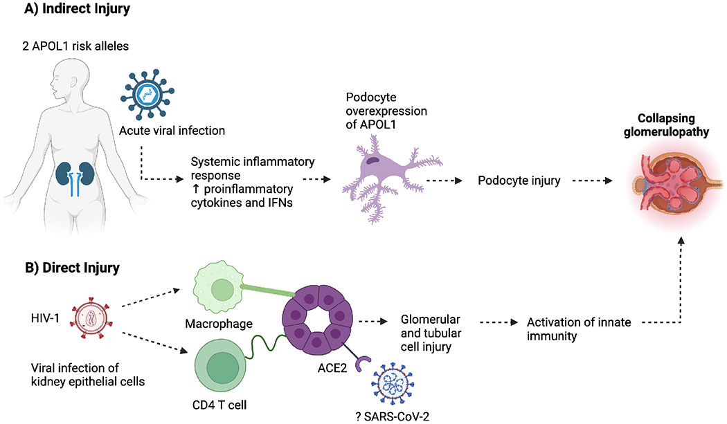
The COVID-19 era has been a reminder to clinicians around the world of the important role that viral infections play in promoting glomerular disease. Several viral infections including human immunodeficiency virus (HIV), severe acute respiratory syndrome coronavirus 2, Epstein-Barr virus, cytomegalovirus, and parvovirus B19 can cause podocyte injury and present with a collapsing glomerulopathy (CG) variant of focal segmental glomerulosclerosis or minimal change disease. CG associated with COVID-19 has been termed COVID-19-associated nephropathy due to its striking resemblance to HIV-associated nephropathy. Host susceptibility is a major determinant of viral infection-associated CG, and the presence of two APOL1 risk variants explains most of the racial predilection to viral-associated CG observed in individuals of African ancestry. Interactions between APOL1 risk variants, viral genes, and the systemic inflammatory response to viral infection all contribute to kidney injury. This review will summarize our current knowledge of viral infection-associated CG, focusing primarily on the clinical presentation, histological features, mechanisms, and disease course of HIV-associated nephropathy and COVID-19-associated nephropathy.

Keywords: COVID-19; Collapsing glomerulopathy; HIV; Podocytopathies; Viruses.


Figures

Figure 1.
Figure 1.. Representative photomicrograph displaying the features of collapsing glomerulopathy
A glomerulus showing the features of collapsing glomerulopathy in a kidney biopsy. This glomerulus displays global collapse and retraction of the glomerular tuft, wrinkled glomerular basement membranes with loss of patency of glomerular capillaries, and both hypertrophy and hyperplasia of the overlying glomerular epithelial cells, some of which contain intracytoplasmic protein resorption droplets (Jones methenamine silver stain). [Original magnification ×400].
Figure 2.
Figure 2.. Photomicrograph displaying a tubular microcyst, a common finding in collapsing glomerulopathy.
This light microscopy image shows dilation of renal tubules by abundant proteinaceous material and accompanying acute tubular injury, manifested by cytoplasmic vacuolization and epithelial simplification in the lining tubular epithelial cells. This is a common tubulointerstitial finding in collapsing glomerulopathy. In fact, this can be a clue to the pathologist that the biopsy may contain a lesion of glomerular collapse, or that such lesion may be present in the patient’s kidney, but not present in the material taken at biopsy. (Hematoxylin & eosin stain) [Original magnification 20x].
Figure 3.
Figure 3.. Electron micrograph showing endothelial cell tubuloreticular inclusions (TRIs).
Two (2) tubuloreticular inclusions (TRIs) [arrows] are identifiable within the cytoplasm of an endothelial cell. They are recognizable as collections of electron dense cytoplasmic elements arranged in a characteristic fashion. These structures are seen in the setting of high interferon states and systemic inflammation. Some authors have termed these structures “interferon footprints”. [JEOL Electron Microscope (Original magnification 80,000x)].
Figure 4.
Figure 4.. Mechanisms of Viral Infection-Associated Collapsing Glomerulopathy
Viral infection-associated collapsing glomerulopathy may occur due to direct and indirect effects of viral infection. A) Indirectly, viral infection results in systemic inflammation and release of proinflammatory cytokines and interferons. Interferon leads to upregulation of APOL1 in podocytes and in individuals who carry 2 APOL1 renal risk variants, this leads to podocyte injury. B) Viruses may also promote injury of kidney cells through direct infection. HIV directly infects kidney tubular epithelial cells and podocytes through cellular contact with infected macrophages and T cells. Viral infection of kidney epithelial cells leads to injury and activation of innate immunity responses and interferon signaling.

Similar articles

Cited by

References

    1. United Nations Programme on HIV and AIDS. Global HIV & AIDS statistics – fact sheet. Available from: https://www.unaids.org/en/resources/fact-sheet. Accessed March 10, 2023
    1. Pardo V, Aldana M, Colton RM, et al. Glomerular lesions in the acquired immunodeficiency syndrome. Ann Intern Med 1984; 101: 429–434. - PubMed
    1. Rao TK, Filippone EJ, Nicastri AD, et al. Associated focal and segmental glomerulosclerosis in the acquired immunodeficiency syndrome. N Engl J Med 1984; 310: 669–673. - PubMed
    1. Genovese G, Tonna SJ, Knob AU, et al. A risk allele for focal segmental glomerulosclerosis in African Americans is located within a region containing APOL1 and MYH9. Kidney Int 2010; 78: 698–704. - PMC - PubMed
    1. Limou S, Nelson GW, Kopp JB, et al. APOL1 kidney risk alleles: population genetics and disease associations. Adv Chronic Kidney Dis 2014; 21: 426–433. - PMC - PubMed

MeSH terms