. 2024 Jul 31;15(7):544.
doi: 10.1038/s41419-024-06934-w.

Eg5 UFMylation promotes spindle organization during mitosis

Affiliations

Eg5 UFMylation promotes spindle organization during mitosis

Guangxu Li et al. Cell Death Dis. .

Abstract

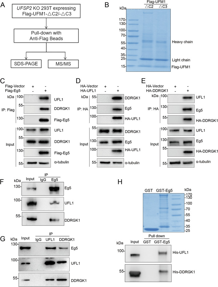
UFMylation is a highly conserved ubiquitin-like post-translational modification that catalyzes the covalent linkage of UFM1 to its target proteins. This modification plays a critical role in the maintenance of endoplasmic reticulum proteostasis, DNA damage response, autophagy, and transcriptional regulation. Mutations in UFM1, as well as in its specific E1 enzyme UBA5 and E2 enzyme UFC1, have been genetically linked to microcephaly. Our previous research unveiled the important role of UFMylation in regulating mitosis. However, the underlying mechanisms have remained unclear due to the limited identification of substrates. In this study, we identified Eg5, a motor protein crucial for mitotic spindle assembly and maintenance, as a novel substrate for UFMylation and identified Lys564 as the crucial UFMylation site. UFMylation did not alter its transcriptional level, phosphorylation level, or protein stability, but affected the mono-ubiquitination of Eg5. During mitosis, Eg5 and UFM1 co-localize at the centrosome and spindle apparatus, and defective UFMylation leads to diminished spindle localization of Eg5. Notably, the UFMylation-defective Eg5 mutant (K564R) exhibited shorter spindles, metaphase arrest, spindle checkpoint activation, and a failure of cell division in HeLa cells. Overall, Eg5 UFMylation is essential for proper spindle organization, mitotic progression, and cell proliferation.


Conflict of interest statement

The authors declare no competing interests.

Figures

Fig. 1
Fig. 1. Eg5 interacts with UFL1 and DDRGK1.
A Strategy for identification of targets for UFMylation. B Proteins eluted from anti-Flag M2 beads were subjected to SDS-PAGE followed by coomassie blue staining. CE Flag-Eg5, HA-UFL1, or HA-DDRGK1 were expressed in HEK293T cells, respectively. Cell lysates were subjected to immunoprecipitation with anti-Flag or anti-HA beads followed by western blot analysis with the indicated antibodies. F, G HEK293T cell lysates were subject to immunoprecipitation with anti-Eg5, anti-UFL1, or anti-DDRGK1 antibodies followed by western blot analysis with the indicated antibodies. IgG was used as a control. H In vitro binding assay. Purified His-UFL1 and His-DDRGK1 were incubated with GST or GST-tagged Eg5, followed by GST pulldown assay. The samples were then subjected to western blot analysis with anti-His antibody.
Fig. 2
Fig. 2. Eg5 is a target substrate for UFMylation.
A Eg5 is UFMylated in vivo. UFMylation system components (HA-UBA5, HA-UFC1, HA-UFL1, HA-DDRGK1, and HA-UFM1) and Flag-Eg5 were expressed in HEK293T cells. Cell lysates were subjected to immunoprecipitation with anti-Flag beads followed by western blot analysis with the indicated antibodies. B UBA5, UFC1, UFL1, and DDRGK1 are required for Eg5 UFMylation. Flag-Eg5 and HA-UFM1-ΔC2 were expressed in UFSP2 knockout HEK293T cells with si-NC, si-UBA5, si-UFC1, si-UFL1, or si-DDRGK1, respectively. Cell lysates were subjected to the UFMylation assay. C Bacterially produced UFMylation components (His-UBA5, His-UFC1, His-UFL1, and His-UFM1-ΔC2) were subjected to Coomassie brilliant blue or western blot analysis with anti-His antibody. D Eg5 is UFMylated in vitro. Purified UFMylation components and GST-Eg5 were incubated in the UFMylation buffer. The reaction was terminated by adding an SDS sample buffer, and the samples were subjected to a western blot with the indicated antibodies.
Fig. 3
Fig. 3. K564 is the essential UFMylation site in Eg5.
A, B Identification of UFMylation region. A series of deletion constructs of Flag-Eg5 were generated as indicated, and expressed in UFSP2 knockout HEK293T cells with HA-UFM1-ΔC2. Cell lysates were subjected to the UFMylation assay. C Identification of the UFMylation site (s). The six Lys residues in the amino acid sequence of 552–641 were replaced by Arg, respectively, and the UFMylation assay was performed in UFSP2 knockout HEK293T cells. D In vitro UFMylation assay of Eg5 and its mutants, as described in Fig. 2D.
Fig. 4
Fig. 4. UFMylation is required for Eg5 monoubiquitination.
A Eg5 stability was examined by western blot in HeLa cells with UFL1 or DDRGK1 knockdown. The cells were treated with 100 µg ml−1 cycloheximide (CHX) for the indicated times, and the Eg5 protein levels were quantified. B Total mRNA was extracted from HeLa cells transfected with the indicated siRNAs, and quantitative real-time RT–PCR assays were performed. C The phosphorylation of Eg5 at Thr926 in HeLa cells transfected with the indicated siRNAs was determined by western blot analysis. D HA-Ub and Flag-Eg5 were expressed in HeLa cells with si-NC, si-UFL1, or si-DDRGK1, respectively. Cell lysates were subjected to immunoprecipitation with anti-Flag beads followed by western blot analysis with the indicated antibodies. The mean ± SD from three independent experiments was shown. The P values were determined by one-way ANOVA. ns, not significant, *P < 0.05; **P < 0.01; ***P < 0.001.
Fig. 5
Fig. 5. Co-localization between UFM1 and Eg5 at the centrosome and spindle.
A The localization of UFM1 and Eg5 was detected by immunofluorescence staining using anti-UFM1 and anti-Eg5 antibodies in HeLa cells at interphase, prophase, metaphase, anaphase, and telophase. The cell nuclei were stained with DAPI. Scale bar, 5 μm. B HeLa cells transfected with HA-UFM1 and Flag-Eg5 were treated with DMSO, Hydroxyurea, and Nocodazole, respectively. The cell lysates were then subjected to a UFMylation assay. The mean ± SD from three independent experiments was shown. The P values were determined by one-way ANOVA. **P < 0.01; ***P < 0.001.
Fig. 6
Fig. 6. Defective Eg5 UFMylation impairs the distribution of Eg5 on the mitotic spindle.
A The localization of Eg5 at the centrosome was detected by immunofluorescence staining using anti-Eg5 and anti-γ-tubulin in HeLa cells. Scale bar, 5 μm. The mean ± SD from at least 45 mitotic cells was shown. The P values were determined by one-way ANOVA. ns, not significant. B Sum intensity of Eg5 in the prophase centrosome region. C The localization of Eg5 at the spindle was detected by immunofluorescence staining using anti-Eg5 and anti-α-tubulin in HeLa cells. DF Sum intensity of Eg5, spindle area, and mean intensity of Eg5 in the metaphase spindle region. Scale bar, 5 μm. The mean ± SD from at least 80 mitotic cells was shown. The P values were determined by one-way ANOVA. *P < 0.05; **P < 0.01; ***P < 0.001.
Fig. 7
Fig. 7. Eg5 UFMylation is required for the spindle assembly and cell proliferation.
HeLa cells were transfected with indicated siRNAs or plasmids. A The phenotypes of the mitotic spindle (monopole, multipole, or disorganized) were determined by immunofluorescence staining using anti-α-tubulin and DAPI in HeLa cells. Transfected cells were identified by Flag positivity. The percent of mitotic phenotypes was calculated. Scale bar, 5 μm. The mean ± SD from three independent experiments, with 100 cells per experimental group, was shown. The P values were determined by two-way ANOVA. ***P < 0.001. B Cell lysates were subjected to western blot analysis with the indicated antibodies. The mean ± SD from three independent experiments was shown. The P values were determined by one-way ANOVA. ***P < 0.001. C Time-lapse imaging of HeLa cells expressing H2B-mCherry and GFP-Tub was conducted following co-transfection with the indicated siRNAs or plasmids. Time was shown as min:s. Scale bar, 10 μm. The duration of metaphase was shown: si-NC+Vector, n = 15; si-Eg5 + Eg5-WT, n = 15; si-NC + Eg5-K564R (n = 25, 15 arrested in metaphase, 10 quantified). The P values were determined by one-way ANOVA. ***P < 0.001. D HeLa cells were cultured for 24 h, 36 h, 48 h, and 72 h, then subjected to CCK-8 assays. The mean ± SD from three independent experiments was shown. The P values were determined by one-way ANOVA. **P < 0.01; ***P < 0.001. E Colony formation assay. HeLa cells were cultured and stained with crystal violet. The number of colonies in each condition was counted. The mean ± SD from three independent experiments was shown. The P values were determined by one-way ANOVA. ***P < 0.001.

References

    1. Komatsu M, Chiba T, Tatsumi K, Iemura S, Tanida I, Okazaki N, et al. A novel protein-conjugating system for Ufm1, a ubiquitin-fold modifier. Embo J. 2004;23:1977–86. 10.1038/sj.emboj.7600205 - DOI - PMC - PubMed
    1. Gerakis Y, Quintero M, Li H, Hetz C. The UFMylation system in proteostasis and beyond. Trends Cell Biol. 2019;29:974–86. 10.1016/j.tcb.2019.09.005 - DOI - PMC - PubMed
    1. Banerjee S, Kumar M, Wiener R. Decrypting UFMylation: how proteins are modified with UFM1. Biomolecules. 2020;10:1442. 10.3390/biom10101442 - DOI - PMC - PubMed
    1. Kang SH, Kim GR, Seong M, Baek SH, Seol JH, Bang OS, et al. Two novel ubiquitin-fold modifier 1 (Ufm1)-specific proteases, UfSP1 and UfSP2. J Biol Chem. 2007;282:5256–62. 10.1074/jbc.M610590200 - DOI - PubMed
    1. Bacik JP, Walker JR, Ali M, Schimmer AD, Dhe-Paganon S. Crystal structure of the human ubiquitin-activating enzyme 5 (UBA5) bound to ATP: mechanistic insights into a minimalistic E1 enzyme. J Biol Chem. 2010;285:20273–80. 10.1074/jbc.M110.102921 - DOI - PMC - PubMed

LinkOut - more resources