. 2024 Jul 31;9(1):134.
doi: 10.1038/s41541-024-00903-2.

A chimeric vaccine derived from Australian genotype IV Japanese encephalitis virus protects mice from lethal challenge

Affiliations

A chimeric vaccine derived from Australian genotype IV Japanese encephalitis virus protects mice from lethal challenge

Jessica J Harrison et al. NPJ Vaccines. .

Abstract

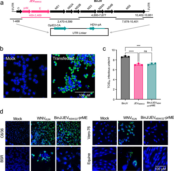
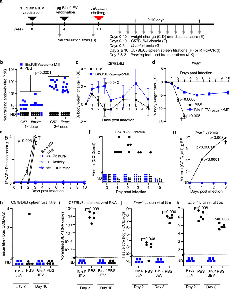
In 2022, a genotype IV (GIV) strain of Japanese encephalitis virus (JEV) caused an unprecedented and widespread outbreak of disease in pigs and humans in Australia. As no veterinary vaccines against JEV are approved in Australia and all current approved human and veterinary vaccines are derived from genotype (G) III JEV strains, we used the recently described insect-specific Binjari virus (BinJV) chimeric flavivirus vaccine technology to produce a JEV GIV vaccine candidate. Herein we describe the production of a chimeric virus displaying the structural prM and E proteins of a JEV GIV isolate obtained from a stillborn piglet (JEVNSW/22) in the genomic backbone of BinJV (BinJ/JEVNSW/22-prME). BinJ/JEVNSW/22-prME was shown to be antigenically indistinguishable from the JEVNSW/22 parental virus by KD analysis and a panel of JEV-reactive monoclonal antibodies in ELISA. BinJ/JEVNSW/22-prME replicated efficiently in C6/36 cells, reaching titres of >107 infectious units/mL - an important attribute for vaccine manufacture. As expected, BinJ/JEVNSW/22-prME failed to replicate in a variety of vertebrate cells lines. When used to immunise mice, the vaccine induced a potent virus neutralising response against JEVNSW/22 and to GII and GIII JEV strains. The BinJ/JEVNSW/22-prME vaccine provided complete protection against lethal challenge with JEVNSW/22, whilst also providing partial protection against viraemia and disease for the related Murray Valley encephalitis virus. Our results demonstrate that BinJ/JEVNSW/22-prME is a promising vaccine candidate against JEV.


Conflict of interest statement

Data included in this publication are based on a patent (WO/2018/176075) on which J.J.H., D.W., H.B-O., R.A.H., and J.H-P. are inventors. The remaining authors declare no competing interests.

Figures

Fig. 1
Fig. 1. Generation and characterisation of the BinJ/JEVNSW/22-prME chimera.
a Schematic of the circular polymerase extension reaction (CPER) strategy to generate infectious DNA of chimeric BinJ/JEVNSW/22-prME. The prME genes of JEVNSW/22 (pink arrows) were inserted into the BinJV backbone (black arrows) (replacing the BinJV prME). OpIE2-CA, a modified Orgyia pseudotsugata multicapsid nucleopolyhedrosis virus immediate-early 2 promoter; HDVr-pA, hepatitis delta virus ribozyme–poly A, with the ribozyme autocleavage providing an authentic 3′ untranslated region (UTR). b IFA analysis of mock (left) and BinJ/JEVNSW/22-prME CPER (right) transfected C6/36 cells fixed 7 days post-transfection and immunolabeled with anti-flavivirus NS1 mAb 4G4 to confirm recovery of replicating virus (green signal). c Growth of chimera was analysed by infecting C6/36 monolayers in triplicate with BinJV, JEVNSW/22 or BinJ/JEVNSW/22-prME at an MOI of 0.1, before titrating on C6/36 monolayers in triplicate and determining viral titres by TCID50. Statistics were performed by one-way ANOVA whereby p = <0.0001 (****) or <0.005 (***). d IFA analysis by confocal microscopy of mock, West Nile virus, Kunjin subtype (WNVKUN, a mammalian cell infection control) and BinJ/JEVNSW/22-prME virus-infected C6/36 cells and mammalian cells at an MOI of 1. Cells were fixed and immunolabelled 5 days after infection. Mammalian cells: BSR (baby hamster kidney), Vero-76 cells (African green monkey kidney) and primary equine dermal fibroblasts. Virus replication was detected with anti-NS1 mAb 4G4 (green signal) and cell nuclei were stained with Hoechst 33342 (blue). Images taken at x40 magnification. Scale bar in (b) and (d) represents 100 μM.
Fig. 2
Fig. 2. BinJ/JEVNSW22-prME vaccine antigen purification.
a BinJ/JEVNSW/22-prME was purified via a potassium tartrate gradient, sedimenting as a wide opalescent blue band (mature virions) or white band (immature virions). b SDS-PAGE (4–12%) analysis of gradient purified BinJ/JEVNSW/22-prME. Flavivirus structural proteins (pre-membrane (prM), capsid (C), membrane (M) and envelope (E)) are indicated. c Negative-stain TEM image of mature (upper panel, x20, 000 magnification) and immature (lower panel, x25, 000 magnification) BinJ/JEVNSW/22-prME virions. Scale bar represents 200 nm. For lower panel, example of mature virion (red arrow) and immature virion (black arrow) indicated. d ELISA curves for indicated purified anti-JEV mAbs using C6/36 fixed cells that had been infected with BinJV-based JEV chimeras GI-V, wild-type WNVKUN and MVEV or mock-infected as the antigen. e Competitive ELISA analysis using saturating concentrations of mAbs listed on the X axis and detection of binding of mAb JV-4H12. f Kd values for binding of mAbs to BinJ/JEVNSW/22-prME and the corresponding JEVNSW/22; each dot represents one mAb (mAbs are described in Table S4). Yellow, E-specific mAbs; orange, E domain II–specific mAbs; green, E domain III–specific mAbs; purple, prM and E-specific mAbs, grey, unspecified prM/E-reactive mAbs. Statistics were performed using Pearson correlations.
Fig. 3
Fig. 3. BinJ/JEVNSW/22-prME vaccination and challenge.
a Timeline of BinJ/JEVNSW/22-prME vaccinations, JEVNSW/22 challenge, and sample/data collections. Silver arrows indicate sample/data collection timepoints. b Serum end-point neutralising antibody titres after one (1st dose) and two (2nd dose) vaccinations. Limit of quantification 1 in 5. (Statistics by Kolmogorov-Smirnov tests). c Mean percent C57BL/6J mouse body weight change relative to day 0 for each mouse (n = 10 per group 0-2 dpi, n = 5 per group 3–10 dpi). Differences only reached significance at 3 dpi, statistics by t-test (significance was not reached on any other day, or ranges of days by repeated measure ANOVA tests). ND not detected. d As for C for Ifnar-/- mice. Statistics by Kolmogorov-Smirnov exact tests (2 and 3 dpi). e Clinical disease scores for posture (circles), activity (crosses) and fur ruffling (triangles) were monitored in Ifnar-/- mice. N as in (c). † Ethically defined end point reached for euthanasia. Statistics by Kolmogorov-Smirnov exact tests (n = 5 per group), p = 0.013 for each disease parameter. f Post-challenge viremia in C57BL/6J mice determined by TCID50 assays. Viremia for individual mice at each time point. Limit of detection was 2 log10TCID50/ml. ND not detected. In the PBS group no viremia was detected on any day in 3/10 mice. Statistics by Kolmogorov-Smirnov exact test for 1–4 dpi (n = 30 serum samples per group; 30 vs. 18 ND). G Post-challenge viremia in Ifnar-/- mice (n = 10 per group 0-2 dpi, n = 5 per group 3 dpi). h Tissue titres for C57BL/6J spleens. Limit of detection ≈2.7 log10TCID50/g. i RT-qPCR of JEV RNA in C57BL/6J spleens. JEV RNA copies are normalized to Rpl13a. Limit of detection ≈10-12 JEV/Rpl13a copies. j Spleen and (k) brain tissue titres in Ifnar-/- mice 2 and 3 dpi.
Fig. 4
Fig. 4. BinJ/JEVNSW/22-prME vaccination and immunogenicity studies.
a Timeline for BinJ/JEVNSW/22-prME vaccinations and immunogenicity studies. Serum taken 16 weeks post-vaccination was assessed in immunogenicity studies. b Total Ig response to each of the 5 JEV genotypes. c Total Ig response to each of the 5 JEV genotypes. Plotted mid-point titres (t50) from data displayed in (b). d Neutralising antibody titres against WT GII (Fu), GIII (Nakayama; SA-14) and GIV (NSW/22). mAb BJ-6E6 was used as a control to ensure sufficient antigen was present. NS not significant.
Fig. 5
Fig. 5. BinJ/JEVNSW/22-prME provides partial cross protection against MVEVTC123130 in Ifnar-/- mice.
a Timeline of BinJ/JEVNSW/22-prME vaccinations, MVEV challenge, and sample/data collections. b Serum end point neutralizing antibody titers against either JEVNSW/22 or MVEV after two vaccinations. Limit of quantification 1 in 5. (Statistics by Wilcoxon matched-pairs signed rank tests). c Mean percent mouse body weight change relative to day 0 for each mouse (n = 6 per group). Statistics by repeated measure ANOVA for 1 to 4 dpi (p = 0.008). d Clinical disease scores for posture (circles), activity (crosses) and fur ruffling were monitored in Ifnar-/- mice. n = 6 PBS injected, n = 12 BinJ/JEVNSW/22-prME vaccinated until 4 dpi, n = 6 BinJ/JEVNSW/22-prME vaccinated until humane endpoint reached. † Ethically defined end point reached for euthanasia. Statistics by Kolmogorov-Smirnov exact tests (n = 6 PBS vaccinated, n = 12 BinJ/JEVNSW/22-prME vaccinated on 4 dpi), p < 0.05 for each disease parameter. e % survival; time until mice reached ethically defined end points for euthanasia, n = 6 per group. Significance by log rank statistic. f Post-challenge viremia in Ifnar-/- mice determined by TCID50 assays. Viremia for individual mice at each time point. Limit of detection was 2 log10TCID50/ml. ND not detected. In the BinJ/JEVNSW/22-prME vaccinated group no viremia was detected on any day in 4/12 mice (blue square with red boarder). Statistics by Kolmogorov-Smirnov exact test on 2 dpi (n = 6 PBS injected, n = 12 BinJ/JEVNSW/22-prME vaccinated).

References

    1. Halstead, S., Hills, S. & Dubischar, K. In Plotkin’s Vaccines (eds Plotkin, S. A., Offit, P. A., Orenstien, W. A., & Edwards, K. M.) 511-548e512 (Elsevier, 2018).
    1. Pierson, T. & Diamond, M. In Fields Virology Vol. 1 (eds Knipe, D. M. et al.) Ch. 26 (Lippincott, Williams and Wilkins, 2013).
    1. Gao, X. et al. Changing geographic distribution of Japanese encephalitis virus genotypes, 1935-2017. Vector Borne Zoonotic Dis19, 35–44 (2019). 10.1089/vbz.2018.2291 - DOI - PubMed
    1. Hanna, J. N. et al. An outbreak of Japanese encephalitis in the Torres Strait, Australia, 1995. Med. J. Aust.165, 256–260 (1996). 10.5694/j.1326-5377.1996.tb124960.x - DOI - PubMed
    1. Ritchie, S. A. & Rochester, W. Wind-blown mosquitoes and introduction of Japanese encephalitis into Australia. Emerg. Infect. Dis.7, 900–903 (2001). 10.3201/eid0705.017524 - DOI - PMC - PubMed

LinkOut - more resources