. 2024 Jul 31;7(1):924.
doi: 10.1038/s42003-024-06632-5.

MiR-199a-3p regulates HCT-8 cell autophagy and apoptosis in response to Cryptosporidium parvum infection by targeting MTOR

Affiliations

MiR-199a-3p regulates HCT-8 cell autophagy and apoptosis in response to Cryptosporidium parvum infection by targeting MTOR

Shanbo Wu et al. Commun Biol. .

Abstract

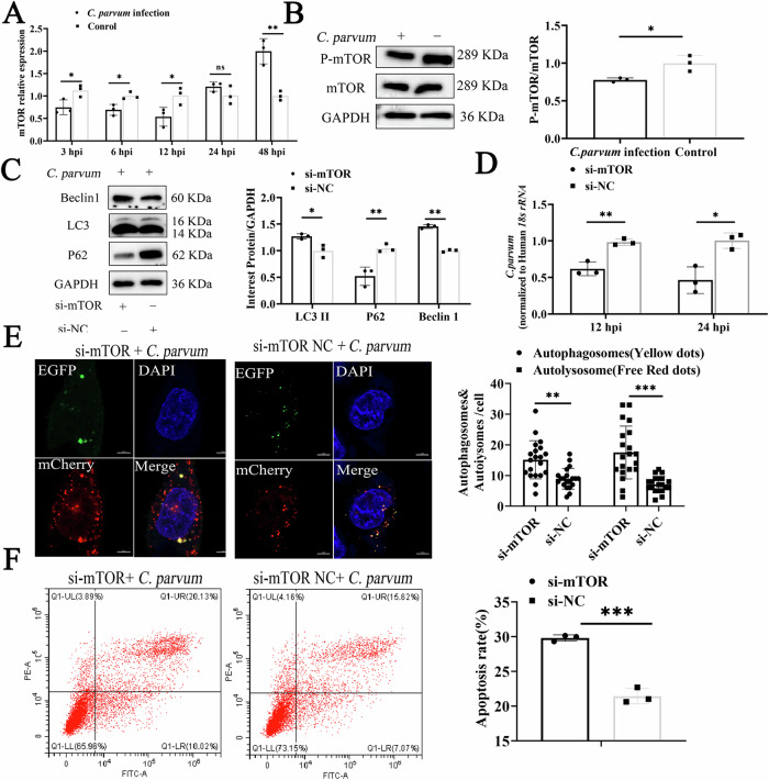
The microRNAs (miRNAs) of their hosts play an important role in regulating both the innate and adaptive immune responses to Cryptosporidium parvum infection. The mechanisms of autophagy and apoptosis are important components of the defense system against C. parvum infection. In this study, we investigate the role of miRNA-199a-3p in regulating MTOR-mediated autophagy and apoptosis in HCT-8 cells induced by C. parvum. The expression of miR-199a-3p increased at 3, 6 and 12 hours postinfection (hpi) but decreased at 24 and 48 hpi. The upregulation of miR-199a-3p promoted autophagy and apoptosis and limited the parasite burden in HCT-8 cells after C. parvum infection. The downregulation of miR-199a-3p inhibited the autophagy and apoptosis induced by C. parvum and enhanced the parasite burden in HCT-8 cells. A luciferase reporter showed that MTOR was a target gene of miR-199a-3p. Suppressed expression of MTOR by small interfering RNA (siRNA) promoted autophagy and apoptosis and limited C. parvum burden in HCT-8 cells. Co-transfection with miR-199a-3p inhibitor or si-mTOR revealed that miR-199a-3p regulates autophagy and apoptosis in HCT-8 cells through MTOR, to resist C. parvum infection. In conclusion, intestinal epithelial cells defend against C. parvum infection by regulating their autophagy and apoptosis through the miR-199a-3p-MTOR axis.


Conflict of interest statement

The authors declare no competing interests.

Figures

Fig. 1
Fig. 1. Complete autophagy of host cells inhibited the intracellular proliferation of Cryptosporidium parvum.
A Expression levels of autophagy-related proteins LC3, P62, and Beclin 1 in HCT-8 cells at different time points after C. parvum sporozoites infection, detected with western blotting. The protein levels of LC3-II, P62, and Beclin 1 relative to GAPDH levels were determined with densitometry(n  =  3 biologically independent samples). B Cells were transfected with plasmid stably expressing EGFP-LC3 and then exposed to equal numbers of C. parvum sporozoites for 12 h. C. parvum were stained with Pan Cp followed by goat anti-mouse IgG Alexa Fluor 594, and the nucleus was stained with DAPI. The spatial relationship between C. parvum and autophagosome was observed by confocal microscopy (red dots: C. parvum; green dots: autophagosome, Scale bars, 2 µm). C HCT-8 cells were pretreated with 500 nM Rapamycin (Rap) or 30 µM Chloroquine (CQ) for 4 h, and then infected with C. parvum sporozoites for 12 h. The expression of LC3 and P62 in the cell pellets was determined with western blotting(n  =  3 biologically independent samples). D Cells were transfected with plasmid stably expressing mCherry-GFP-LC3 and then exposed to equal numbers of C. parvum sporozoites for 12 h (n = 3 biologically independent samples). The autophagic flux was evaluated with confocal microscopy. Numbers of yellow and free red dots in each group cell were quantified (free red dots: autophagic lysosomes; yellow dots: autophagosome, Scale bar, 5 µm). E Effects of 500 nM Rap and 30 µM CQ on the propagation of C. parvum in HCT-8 cells. HCT-8 cells were pretreated with 500 nM Rap or 30 µM CQ for 4 h and infected with C. parvum sporozoites, which were quantified with RT–qPCR assays at 12 and 24 hpi(n  =  3 biologically independent samples). All data are presented as the means ± SD of three independent experiments. Different groups were compared with a t test. (*P < 0.05, **P < 0.01, ***P < 0.001).
Fig. 2
Fig. 2. MiR-199a-3p regulates autophagy and apoptosis in cells to limit the C.
parvum load after infection. A Expression levels of miR-199a-3p in HCT-8 cells at different times after C. parvum sporozoites infection, determined with RT-qPCR (n  =  3 biologically independent samples). B Expression efficiency of miR-199a-3p in HCT-8 cells transfected with miR-199a-3p mimic or miR-199a-3p inhibitor for 24 h, determined with RT-qPCR. C Effect of miR-199a-3p on autophagy of cells infected with C. parvum. Cells were transfected with miR-199a-3p mimic or miR-199a-3p inhibitor for 24 h and then exposed to equal numbers of C. parvum sporozoites for 12 h. Expression levels of autophagy-related proteins LC3, P62, and Beclin 1 were determined with western blotting. Protein levels of LC3-II, P62, and Beclin 1 relative to GAPDH levels were determined with densitometry(n  =  3 biologically independent samples). D Effect of miR-199a-3p on autophagic flux in C. parvum-infected cells. Cells were cotransfected with miR-199a-3p mimic or miR-199a-3p inhibitor and a plasmid encoding mCherry–EGFP–LC3 for 24 h, and then exposed to equal numbers of C. parvum sporozoites for 12 h (n = 3 biologically independent samples). The autophagic flux was evaluated with confocal microscopy. Numbers of yellow and free red dots in each group cell were quantified (free red dots: autophagic lysosomes; yellow dots: autophagosome, Scale bar, 5 µm). E Effect of miR-199a-3p on apoptosis of cells infected with C. parvum. Cells were transfected with miR-199a-3p mimic or miR-199a-3p inhibitor for 24 h and then exposed to equal numbers of C. parvum sporozoites for 12 h. Cell apoptosis was detected with flow cytometry. The apoptosis rate is the sum of O1-LR and O1-UR (n  =  3 biologically independent samples). F Cells were transfected with miR-199a-3p mimic or miR-199a-3p inhibitor for 24 h and then exposed to equal numbers of C. parvum sporozoites for 2 h to determine the initial attachment and cellular invasion of C. parvum with RT-qPCR. The medium was replaced with fresh medium at 3 hpi infection with C. parvum and the infected cells cultured for a further 12 or 24 h to evaluate the parasite burden after invasion(n  =  3 biologically independent samples). All data presented are the means ± SD of three independent experiments. Different groups were compared with a t test (*P < 0.05, **P < 0.01, ***P < 0.001).
Fig. 3
Fig. 3. MiR-199a-3p regulates the expression of MTOR.
A Binding sites of miR-199a-3p on MTOR predicted with TargetScan. B Binding of miR-199a-3p to MTOR was detected with a dual-luciferase assay in HCT-8 cells cotransfected with miR-199a-3p or miR-199a-3p NC and pmirGLO–MTOR-WT or pmirGLO–MTOR-MUT for 48 h (n  =  5 biologically independent samples). C, D Cells were transfected with miR-199a-3p mimic or miR-199a-3p inhibitor for 24 h and then exposed to equal numbers of C. parvum sporozoites for 12 h. MTOR mRNA and protein expression levels were detected with RT–qPCR and western blotting, respectively. Densitometric levels of MTOR protein signals and mRNA levels were quantified and expressed relative to GAPDH (n  =  3 biologically independent samples). All data are presented as the means ± SD of three independent experiments. Different groups were compared with a t test (*P < 0.05, **P < 0.01, ***P < 0.001).
Fig. 4
Fig. 4. MTOR is involved in regulating the autophagy and apoptosis of HCT-8 cells induced by C. parvum.
A MTOR mRNA expression was detected with RT–qPCR(n  =  3 biologically independent samples). B Levels of phosphorylated MTOR (p-MTOR) and total MTOR were analyzed with western blotting 12 h after HCT-8 cells were infected with C. parvum sporozoites. Densitometry was used to compare the relative levels of p-MTOR protein and total MTOR protein, and the data were normalized to GAPDH(n  =  3 biologically independent samples). C Cells were transfected with si-mTOR or si-NC for 24 h and then exposed to equal numbers of C. parvum sporozoites for 12 h. Expression levels of autophagy-related proteins LC3, P62, and Beclin 1 were determined with western blotting. Protein levels of LC3-II, P62, and Beclin 1 were determined relative to GAPDH with densitometry(n  =  3 biologically independent samples). D Cells were transfected with si-MTOR or si-NC for 24 h and then exposed to equal numbers of C. parvum sporozoites for 3 h before the medium was replaced with fresh medium. The infected cells were cultured for 12 or 24 h to evaluate the parasite burden with RT–qPCR after cell invasion(n  =  3 biologically independent samples). E Cells were cotransfected with si-MTOR or si-NC and plasmid encoding mCherry–EGFP–LC3 for 24 h, and then exposed to equal numbers of C. parvum sporozoites for 12 h(n  =  3 biologically independent samples). The autophagic flux was evaluated with confocal microscopy. Numbers of yellow and free red dots in each group cell were quantified (free red dots: autophagic lysosomes; yellow dots: autophagosome, Scale bar, 5 µm). F Cells were transfected with si-MTOR or si-NC for 24 h and then exposed to equal numbers of C. parvum sporozoites for 12 h. Cell apoptosis was then detected with flow cytometry. The apoptosis rate is the sum of O1-LR and O1-UR(n = 3 biologically independent samples). All data are presented as the means ± SD of three independent experiments. Different groups were compared with a t test (*P < 0.05, **P < 0.01, ***P < 0.001).
Fig. 5
Fig. 5. MiR-199a-3p-regulated cell autophagy and apoptosis reduced the C.
parvum burden in HCT-8 cells by targeting MTOR. A Cells were cotransfected with miR-199a-3p inhibitor or miR-199a-3p inhibitor NC and si-mTOR or si-NC for 24 h, and then exposed to equal numbers of C. parvum sporozoites for 12 h. Expression levels of autophagy-related proteins LC3, P62, and Beclin 1 were determined with western blotting. Protein levels of LC3II, P62, and Beclin 1 were determined relative to GAPDH protein levels with densitometry(n = 3 biologically independent samples). B Cells were cotransfected with miR-199a-3p inhibitor or miR-199a-3p inhibitor NC and si-MTOR or si-NC for 24 h, and then exposed to equal numbers of C. parvum sporozoites for 12 h. Cell apoptosis was then detected with flow cytometry. The apoptosis rate is the sum of O1-LR and O1-UR(n = 3 biologically independent samples). C Expression efficiency of miR-199a-3p in HCT-8 cells cotransfected with miR-199a-3p inhibitor and si-MTOR for 24 h, determined with RT–qPCR. D Cells cotransfected with miR-199a-3p inhibitor or miR-199a-3p inhibitor NC and si-MTOR or si-NC for 24 h were then exposed to equal numbers of C. parvum sporozoites for 3 h. The medium was then replaced with fresh medium. The infected cells were cultured for 12 or 24 h to evaluate the parasite burden after cell invasion, with RT-qPCR(n = 3 biologically independent samples). All data are presented as the means ± SD of three independent experiments. Different groups were compared with a t test (*P < 0.05, **P < 0.01, ***P < 0.001).

References

    1. O’Hara, S. P. & Chen, X. M. The cell biology of Cryptosporidium infection. Microbes Infect.13, 721–730 (2011). 10.1016/j.micinf.2011.03.008 - DOI - PMC - PubMed
    1. Feng, Y., Ryan, U. M. & Xiao, L. Genetic diversity and population structure of cryptosporidium. Trends Parasitol.34, 997–1011 (2018). 10.1016/j.pt.2018.07.009 - DOI - PubMed
    1. Ryan, U. M., Feng, Y., Fayer, R. & Xiao, L. Taxonomy and molecular epidemiology of Cryptosporidium and Giardia a 50 year perspective (1971-2021). Int. J. Parasitol.51, 1099–1119 (2021). 10.1016/j.ijpara.2021.08.007 - DOI - PubMed
    1. McCole, D. F., Eckmann, L., Laurent, F. & Kagnoff, M. F. Intestinal epithelial cell apoptosis following Cryptosporidium parvum infection. Infect. Immun.68, 1710–1713 (2000). 10.1128/IAI.68.3.1710-1713.2000 - DOI - PMC - PubMed
    1. Deng, M., Rutherford, M. S. & Abrahamsen, M. S. Host intestinal epithelial response to Cryptosporidium parvum. Adv. drug Deliv. Rev.56, 869–884 (2004). 10.1016/j.addr.2003.10.034 - DOI - PubMed

Publication types

MeSH terms