

The protective effect of imatinib against pancreatic β-cell apoptosis induced by dexamethasone via increased GSTP1 expression and reduced oxidative stress
- PMID: 39085384
- PMCID: PMC11291718
- DOI: 10.1038/s41598-024-68429-0
The protective effect of imatinib against pancreatic β-cell apoptosis induced by dexamethasone via increased GSTP1 expression and reduced oxidative stress
Abstract
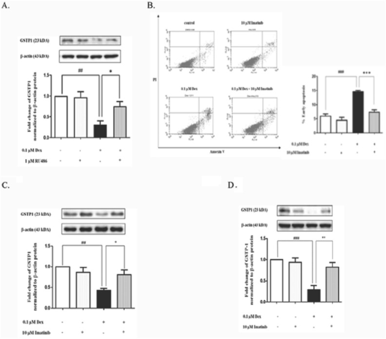
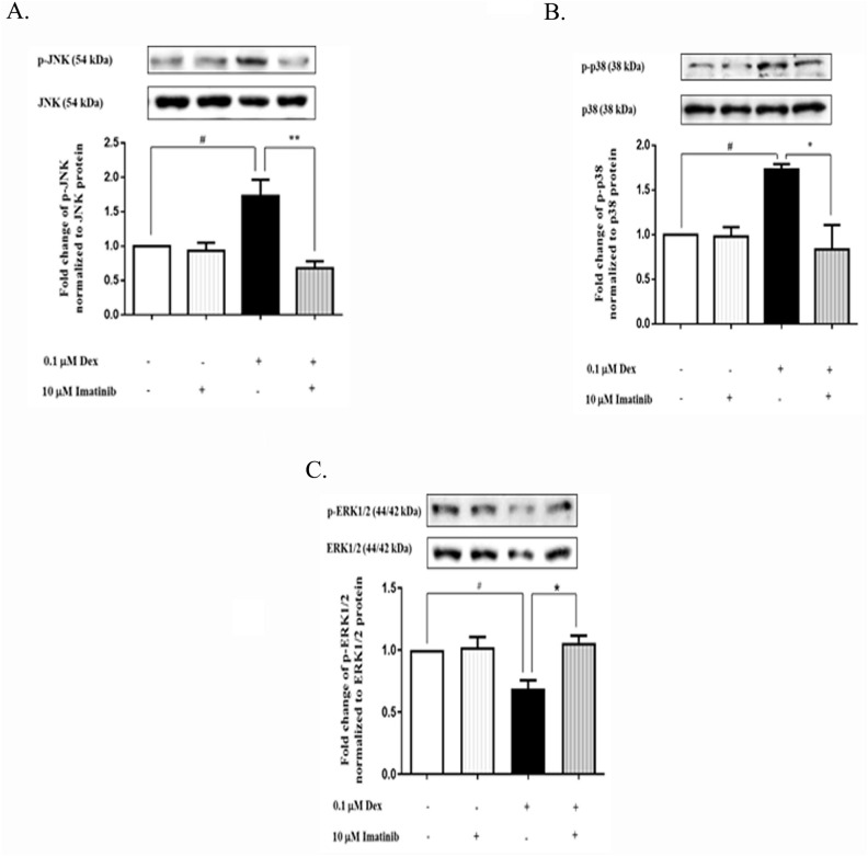
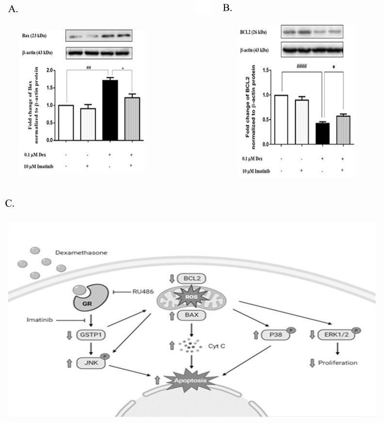
Glucocorticoids (GCs) are known to stimulate pancreatic beta (β)-cell apoptosis via several mechanisms, including oxidative stress. Our previous study suggested an increase in dexamethasone-induced pancreatic β-cell apoptosis via a reduction of glutathione S-transferase P1 (GSTP1), which is an antioxidant enzyme. Imatinib, which is a tyrosine kinase inhibitor, also exerts antioxidant effect. This study aims to test our hypothesis that imatinib would prevent pancreatic β-cell apoptosis induced by dexamethasone via increased GSTP1 expression and reduced oxidative stress. Our results revealed that dexamethasone significantly increased apoptosis in INS-1 cells when compared to the control, and that imatinib significantly decreased INS-1 cell apoptosis induced by dexamethasone. Moreover, dexamethasone significantly increased superoxide production in INS-1 cells when compared to the control; however, imatinib, when combined with dexamethasone, significantly reduced superoxide production in INS-1 cells. Dexamethasone significantly decreased GSTP1, p-ERK1/2, and BCL2 protein expression, but significantly increased p-JNK, p-p38, and BAX protein expression in INS-1 cells-all compared to control. Importantly, imatinib significantly ameliorated the effect of dexamethasone on the expression of GSTP1, p-ERK1/2, p-JNK, p-p38 MAPK, BAX, and BCL2. Furthermore-6-(7-nitro-2,1,3-benzoxadiazol-4-ylthio) hexanol (NBDHEX), which is a GSTP1 inhibitor, neutralized the protective effect of imatinib against pancreatic β-cell apoptosis induced by dexamethasone. In conclusion, imatinib decreases pancreatic β-cell apoptosis induced by dexamethasone via increased GSTP1 expression and reduced oxidative stress.
Keywords: Apoptosis; Dexamethasone; GSTP1; Glucocorticoid receptor; Pancreatic β-cells.
© 2024. The Author(s).
Conflict of interest statement
The authors declare no competing interests.
Figures

Similar articles
-
Imatinib prevents dexamethasone-induced pancreatic β-cell apoptosis via decreased TRAIL and DR5.J Cell Biochem. 2023 Sep;124(9):1309-1323. doi: 10.1002/jcb.30450. Epub 2023 Aug 9. J Cell Biochem. 2023. PMID: 37555250
-
Targeting GSTP1-1 induces JNK activation and leads to apoptosis in cisplatin-sensitive and -resistant human osteosarcoma cell lines.Mol Biosyst. 2012 Apr;8(4):994-1006. doi: 10.1039/c1mb05295k. Epub 2011 Nov 9. Mol Biosyst. 2012. PMID: 22068640
-
GSK-3β mediates dexamethasone-induced pancreatic β cell apoptosis.Life Sci. 2016 Jan 1;144:1-7. doi: 10.1016/j.lfs.2015.11.017. Epub 2015 Nov 25. Life Sci. 2016. PMID: 26606859 Free PMC article.
-
Modulatory role of imatinib mesylate on pancreatic β-cells' secretory functions in an STZ rat model of diabetes mellitus.Chem Biol Interact. 2020 Sep 1;328:109197. doi: 10.1016/j.cbi.2020.109197. Epub 2020 Jul 22. Chem Biol Interact. 2020. PMID: 32710900
-
6-(7-nitro-2,1,3-benzoxadiazol-4-ylthio) hexanol: a promising new anticancer compound.Biosci Rep. 2018 Feb 13;38(1):BSR20171440. doi: 10.1042/BSR20171440. Print 2018 Feb 28. Biosci Rep. 2018. PMID: 29358310 Free PMC article. Review.
Cited by
-
METTL3 inhibits primed-to-naïve transition of pluripotent stem cells through m6A-YTHDF2-pluripotency/Gstp1 mRNA degradation axis.Cell Regen. 2025 May 27;14(1):19. doi: 10.1186/s13619-025-00241-1. Cell Regen. 2025. PMID: 40423919 Free PMC article.
References
MeSH terms
Substances
Grants and funding
LinkOut - more resources
Full Text Sources
Research Materials
Miscellaneous
