. 2024 Nov;19(11):1735-1744.
doi: 10.1038/s41565-024-01727-w. Epub 2024 Jul 31.

Polymersomes with splenic avidity target red pulp myeloid cells for cancer immunotherapy

Affiliations

Polymersomes with splenic avidity target red pulp myeloid cells for cancer immunotherapy

Annelies C Wauters et al. Nat Nanotechnol. 2024 Nov.

Abstract

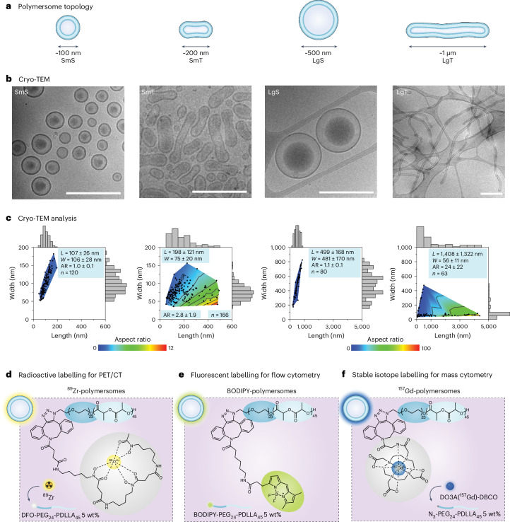
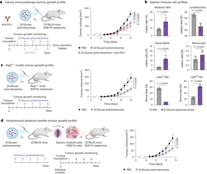
Regulating innate immunity is an emerging approach to improve cancer immunotherapy. Such regulation requires engaging myeloid cells by delivering immunomodulatory compounds to hematopoietic organs, including the spleen. Here we present a polymersome-based nanocarrier with splenic avidity and propensity for red pulp myeloid cell uptake. We characterized the in vivo behaviour of four chemically identical yet topologically different polymersomes by in vivo positron emission tomography imaging and innovative flow and mass cytometry techniques. Upon intravenous administration, relatively large and spherical polymersomes accumulated rapidly in the spleen and efficiently targeted myeloid cells in the splenic red pulp. When loaded with β-glucan, intravenously administered polymersomes significantly reduced tumour growth in a mouse melanoma model. We initiated our nanotherapeutic's clinical translation with a biodistribution study in non-human primates, which revealed that the platform's splenic avidity is preserved across species.


Conflict of interest statement

Competing interests W.J.M.M., L.A.B.J., M.G.N. and Z.A.F. are scientific founders of Trained Therapeutix Discovery. The remaining authors declare no competing interests.

Figures

Fig. 1
Fig. 1. Schematic overview of the study.
a, Polymersomes consist of biodegradable PEG-PDLLA block copolymers assembled into bilayered vesicles. Polymersome topology can be precisely controlled to form spheres and tubes of different sizes (SmS and LgS denote small and large spheres and SmT and LgT denote small and large tubes, respectively). Furthermore, polymersomes are labelled with radionuclides (89Zr), fluorescent dyes (BODIPY) or stable isotopes (157Gd). b, Polymersome in vivo biodistribution was assessed by PET/CT imaging in B16F10 melanoma-bearing C57BL/6 mice upon polymersome administration intravenously (i.v.) via lateral tail vein injection or subcutaneously (s.c.) via footpad injection. Using flow and mass cytometry, we studied polymersome uptake by specific immune cells, including the different splenic compartments, that is, white pulp, red pulp and the marginal zone. c, We loaded polymersomes with β-glucan, applied either as a monotherapy or in combination with anti-PD-1 checkpoint inhibition. d, To study the clinical potential of our nanotherapeutic, we evaluated the biodistribution and biocompatibility of the large spherical β-glucan-polymersomes in NHPs by PET/CT imaging. DC, dendritic cell; MΦ, macrophage; Mo, monocyte. Schematic illustrations of mice, spleens, lymph nodes, syringes, cells, antibodies and NHPs were created with BioRender.com.
Fig. 2
Fig. 2. Characterizing and labelling polymersomes.
a, Schematic representation of the four different polymersome topologies used in this study. b, Representative cryo-TEM micrographs of the four BODIPY-labelled polymersome topologies in their respective dialysis fluids, that is, SmS and LgS, were measured in Milli-Q water and SmT and LgT in 50 mM NaCl. Scale bars, 500 nm. Similar results for polymersome topologies were obtained in three independent experiments. c, Polymersome dimensions determined using the cryo-TEM micrographs. The heatmap indicates the corresponding polymersome aspect ratios. d, Schematic representation of the polymersome radiolabelling strategy. DFO-functionalized polymersomes were formulated by incorporating 5 wt% DFO-PEG24-PDLLA45 followed by incubation with radioactive 89Zr to yield 89Zr-polymersomes. e, Schematic representation of the polymersome fluorescent labelling strategy. BODIPY-polymersomes were formulated by incorporating 5 wt% BODIPY-PEG24-PDLLA45. f, Schematic representation of the polymersome stable isotype labelling strategy. N3-functionalized polymersomes were formulated by incorporating 5 wt% N3-PEG24-PDLLA45. These polymersomes were subsequently incubated with stable 157Gd complexed in DO3A-DBCO to yield 157Gd-polymersomes. All data are represented as mean ± s.d.
Fig. 3
Fig. 3. Polymersome biodistribution and immune cell specificity in the B16F10 melanoma mouse model.
a,b, Schematic overview of 89Zr-polymersome biodistribution studies by PET/CT imaging (a) and BODIPY-polymersome immune cell specificity studies by flow cytometry (b). c, Representative whole-body PET images taken 48 h after subcutaneous administration of 89Zr-polymersomes via footpad injection. d, Biodistribution of subcutaneously administered 89Zr-polymersomes, n = 4 per group. e, Uptake of subcutaneously administered BODIPY-polymersomes in specific immune cell types in the popliteal and iliac lymph nodes, n = 4 per group. f, Representative whole-body PET images taken 48 h after intravenous administration of 89Zr-polymersomes via lateral tail vein injection. g, Biodistribution of intravenously administered 89Zr-polymersomes, n = 4 per group. h, Uptake of intravenously administered BODIPY-polymersomes by immune cells in the spleen and bone marrow, n = 3 per group. Heatmaps of d and g show the average percentage injected dose per gram of tissue (%ID g−1) of the 89Zr-polymersomes per tissue as measured by ex vivo gamma counting. Heatmaps of e and h show the average BODIPY mean fluorescence intensity (MFI) per cell type. i, Schematic overview of 157Gd-polymersome splenocyte specificity study by mass cytometry. j, Uptake of intravenously administered 157Gd-polymersomes by red pulp, marginal zone or white pulp-resident splenocytes. The average 157Gd mean intensity for each cell type, n = 4 per group. BM, bone marrow; cDCs, classical DCs; HSPCs, hematopoietic stem and progenitor cells; ILN, iliac lymph node; Lym, lymphocytes; MZMΦφ, marginal zone macrophages; MMMΦ, marginal metallophilic macrophages; Nφ, neutrophils; NK cells, natural killer cells; PK, pharmacokinetics; PLN, popliteal lymph node; pDCs, plasmacytoid DCs; RPMΦ, red pulp macrophages. Data are presented as mean ± s.e.m. P values were calculated using a two-way ANOVA to compare polymersome topology per splenocyte subset. All P values are two-tailed and P < 0.05 is considered significant. Schematic illustrations of mice, syringes and spleens were created with BioRender.com. Source data
Fig. 4
Fig. 4. β-Glucan-loaded polymersomes modulate splenic myeloid cells and reduce tumour growth in the B16F10 melanoma mouse model.
a, β-Glucan-polymersomes, as a monotherapy or combined with anti-PD-1 checkpoint inhibition, were administered intravenously to B16F10 melanoma-bearing C57BL/6 mice and tumour growth was monitored for 8 days. The tumour growth profiles show that β-glucan-polymersome immunotherapy effectively inhibited tumour growth as a monotherapy and in combination with anti-PD-1 checkpoint therapy, n = 10 per group. b, Relative abundance of splenic myeloid cells, lymphocytes, neutrophils, monocytes, Ly6Clo monocytes and Ly6Chi monocytes in B16F10 melanoma-bearing C57BL/6 mice treated with PBS and β-glucan-polymersomes. The abundance of splenic myeloid cells, including neutrophils and monocytes, was significantly increased in mice treated with β-glucan-polymersomes compared with PBS. The frequency of splenic Ly6Clo monocytes was significantly lower, whereas splenic Ly6Chi monocytes were significantly more abundant in mice treated with β-glucan-polymersomes, n = 8–10 per group. c, β-Glucan-polymersomes were administered intravenously to B16F10 melanoma-bearing Rag1/ mice and tumour growth was monitored for 8 days. Tumour growth profiling revealed that β-glucan-polymersome immunotherapy effectively inhibited tumour growth in lymphocyte-deficient Rag1−/− mice, n = 10 per group. d, Healthy C57BL/6 mice were treated intravenously with β-glucan-polymersomes and splenic myeloid cells were isolated and co-inoculated with B16F10 melanoma cells. Tumour growth was monitored for 8 days. Results show that β-glucan-polymersome-treated myeloid cells inhibited tumour growth when co-inoculated with B16F10 melanoma cells, n = 10 per group. Tumour growth data are presented as mean ± s.e.m. and flow cytometry data are presented as mean ± s.d. P values were calculated using a repeated measures two-way ANOVA to compare tumour growth or an unpaired Mann–Whitney test to compare splenic immune cells. All P values are two-tailed and P < 0.05 is considered significant. i.t., intratumoural. Schematic illustrations of mice, spleens, syringes and cells were created with BioRender.com. Source data
Fig. 5
Fig. 5. β-Glucan-polymersome biodistribution and biocompatibility studies in NHPs confirm nanocarriers’ splenic avidity across different species.
a, Schematic representation of the study. 89Zr-labelled β-glucan-polymersomes were intravenously injected in two NHPs that then underwent dynamic and static PET/CT imaging. b, Dynamic PET/CT images at 1 min, 5 min, 15 min, 30 min and 60 min after injections, showing rapid accumulation of 89Zr-β-glucan-polymersomes in the spleen and liver. c, Quantified uptake of 89Zr-β-glucan-polymersomes in representative organs from images in b, indicating higher accumulation in the spleen than other organs, n = 1. d, PET/CT images at 48 h after injections reveal accumulation in the spleen, liver and bone marrow. e, Quantified uptake of 89Zr-β-glucan-polymersomes in representative organs at 48 h from images in d, n = 2. f, Blood chemistry performed on NHP serum taken before (Pre) and 48 h after (Post) 89Zr-β-glucan-polymersome administration, n = 2. The grey boxes indicate reference values. Alanine transaminase (ALT), aspartate transaminase (AST), creatine and blood urea nitrogen (BUN) levels show no signs of severe toxicity. Schematic illustrations of syringe and NHP were created with BioRender.com.

References

    1. Theobald, M. (ed). Current Immunotherapeutic Strategies in Cancer (Springer, 2020).
    1. Sharma, P. et al. The next decade of immune checkpoint therapy. Cancer Discov.11, 838–857 (2021). - PubMed
    1. Engblom, C., Pfirschke, C. & Pittet, M. J. The role of myeloid cells in cancer therapies. Nat. Rev. Cancer16, 447–462 (2016). - PubMed
    1. Jung, K. et al. Ly6Clo monocytes drive immunosuppression and confer resistance to anti-VEGFR2 cancer therapy. J. Clin. Invest.127, 3039–3051 (2017). - PMC - PubMed
    1. Kubli, S. P., Berger, T., Araujo, D. V., Siu, L. L. & Mak, T. W. Beyond immune checkpoint blockade: emerging immunological strategies. Nat. Rev. Drug Discov.20, 899–919 (2021). - PubMed

MeSH terms

LinkOut - more resources