. 2024 Nov;38(16):3161-3168.
doi: 10.1038/s41433-024-03256-1. Epub 2024 Jul 31.

Effect of baseline fluid localization on visual acuity and prognosis in type 1 macular neovascularization treated with anti-VEGF

Affiliations

Effect of baseline fluid localization on visual acuity and prognosis in type 1 macular neovascularization treated with anti-VEGF

Etienne Gadiollet et al. Eye (Lond). 2024 Nov.

Abstract

Purpose: To assess the prognostic value of subretinal (SRF) and intraretinal fluid (IRF) localizations in type 1 macular neovascularization (MNV) due to age-related macular degeneration (AMD).

Subjects: Eyes were prospectively treated with anti-vascular epithelial growth factor (anti-VEGF) intravitreal injections (IVT) according to a Pro-Re-Nata (PRN) or Treat and Extend (TAE) regimen during 24 months. A total of 211 eyes with treatment-naïve type 1 MNV secondary to AMD were consecutively included. Eyes were divided between 2 groups according to the fluid localization: presence of SRF alone (SRF group), or presence of IRF associated or not with SRF (IRF ± SRF group).

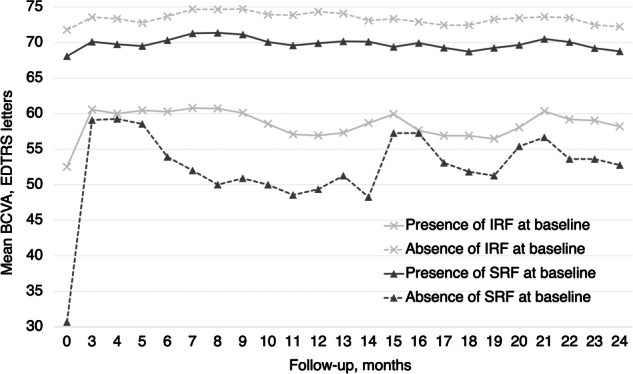
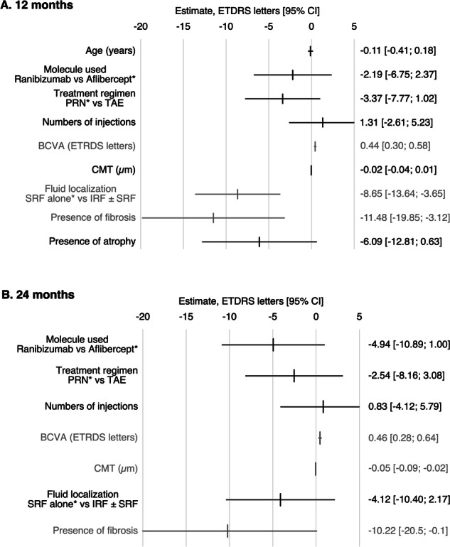
Results: At baseline the mean BCVA was 66.2 letters. SRF was present in 94.8% of eyes, IRF in 30.8%, and both in 25.6%. Data were available for 201 eyes at 12 months, and 157 eyes at 24 months. The presence of IRF at baseline was associated with lower baseline BCVA and significantly lower BCVA at 12 months (p < 0.001) and 24 months (p < 0.001). Eyes with SRF alone displayed better visual outcomes (BCVA at month 12, SRF = 74.3 letters, IRF ± SRF = 56.9 letters). In the presence of baseline IRF, fibrosis (p = 0.03) and atrophy (p < 0.001) were more frequently found at 24 months. In a multivariate model, the presence of baseline IRF was significantly associated with lower BCVA at month 12 but not at month 24.

Conclusion: In type 1 MNV, the presence of baseline IRF was associated with worse visual outcomes compared to SRF alone, and more frequent atrophy and fibrosis.


Conflict of interest statement

The authors declare no competing interests.

Figures

Fig. 1
Fig. 1. BCVA according to the presence of IRF and SRF from baseline to month 24.
BCVA best-corrected visual acuity, ETDRS early treatment diabetic retinopathy study, IRF intraretinal fluid, SRF subretinal fluid.
Fig. 2
Fig. 2. Association between baseline variables and final best corrected visual acuity (BCVA) at 12 and 24 months.
The variables marked with an asterix are the reference variable. A Baseline BCVA, baseline fluid localization and baseline presence of fibrosis are significantly associated with BCVA at 12 months. B Baseline BCVA, the baseline CMT and baseline presence of fibrosis are significantly associated with BCVA at 24 months. BCVA best-corrected visual acuity; CI confidence interval; CMT central macular thickness; ETDRS Early Treatment Diabetic Retinopathy Study; IRF intraretinal fluid; PED pigment epithelium detachment; SRF subretinal fluid.
Fig. 3
Fig. 3. Panel of 2 cases displaying typical evolution with SRF alone and IRF + SRF from baseline to 24 months.
A At baseline, FA displayed multiple drusen associated with ill-defined hyperfluorescence (A1) corresponding to late staining on ICGA (A2) and SRF opposite to a PED on OCT B scans (A3), defining a type 1 MNV. At month 24 (A4), ELM and EZ-IZ complex and RPE remained intact (dotted magnification). B At baseline, FA displayed ill-defined hyperfluorescence (B1) corresponding to a late staining on ICGA (B2) and IRF opposite to a PED on OCT-B scans (B3) defining a type 1 MNV. At 24 months (B4), OCT-B scans displayed ELM disruption, interruption of IZ-EZ complex with alteration of the RPE and hypersignal transmission to the choroid.

References

    1. Congdon N, O'colmain B, Klaver CC, Klein R, Muñoz B, Friedman DS, et al. Causes and prevalence of visual impairment among adults in the United States. Arch Ophthalmol Chic Ill 1960. 2004;122:477–85. 10.1001/archopht.122.4.477. - PubMed
    1. Rosenfeld PJ, Brown DM, Heier JS, Boyer DS, Kaiser PK, Chung CY, et al. Ranibizumab for neovascular age-related macular degeneration. N Engl J Med. 2006;355:1419–31. 10.1056/NEJMoa054481. - PubMed
    1. Schmitz-Valckenberg S, Sahel JA, Danis R, Fleckenstein M, Jaffe GJ, Wolf S, et al. Natural history of geographic atrophy progression secondary to age-related macular degeneration (Geographic Atrophy Progression Study). Ophthalmology. 2016;123:361–8. 10.1016/j.ophtha.2015.09.036. - PubMed
    1. Brown DM, Michels M, Kaiser PK, Heier JS, Sy JP, Ianchulev T, et al. Ranibizumab versus verteporfin photodynamic therapy for neovascular age-related macular degeneration: two-year results of the ANCHOR study. Ophthalmology. 2009;116:57–65.e5. 10.1016/j.ophtha.2008.10.018. - PubMed
    1. Daruich A, Matet A, Moulin A, Kowalczuk L, Nicolas M, Sellam A, et al. Mechanisms of macular edema: beyond the surface. Prog Retin Eye Res. 2018;63:20–68. 10.1016/j.preteyeres.2017.10.006. - PubMed

MeSH terms

LinkOut - more resources