. 2024 Aug;632(8026):850-857.
doi: 10.1038/s41586-024-07749-7. Epub 2024 Jul 31.

Dopamine biases decisions by limiting temporal integration

Affiliations

Dopamine biases decisions by limiting temporal integration

Aditya K Gautham et al. Nature. 2024 Aug.

Abstract

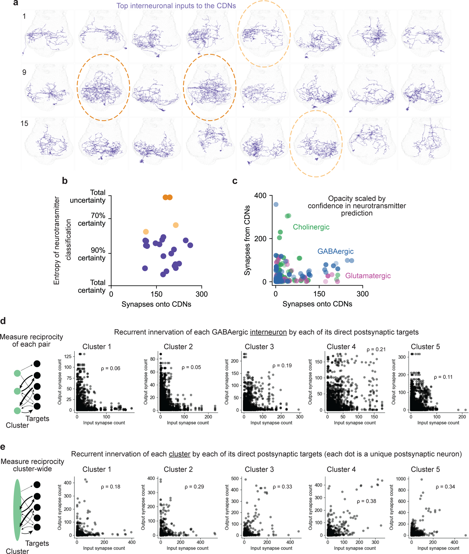
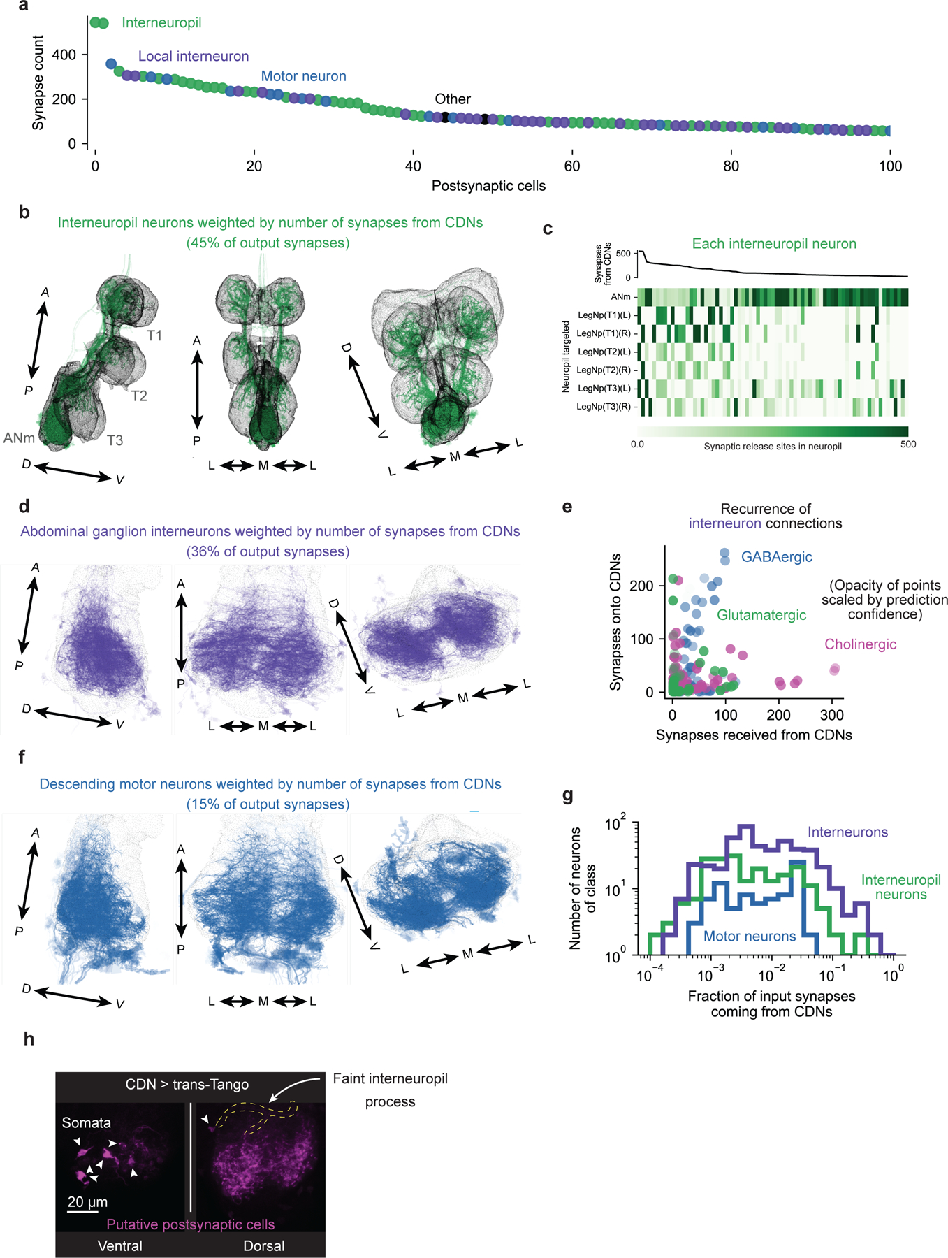
Motivations bias our responses to stimuli, producing behavioural outcomes that match our needs and goals. Here we describe a mechanism behind this phenomenon: adjusting the time over which stimulus-derived information is permitted to accumulate towards a decision. As a Drosophila copulation progresses, the male becomes less likely to continue mating through challenges1-3. We show that a set of copulation decision neurons (CDNs) flexibly integrates information about competing drives to mediate this decision. Early in mating, dopamine signalling restricts CDN integration time by potentiating Ca2+/calmodulin-dependent protein kinase II (CaMKII) activation in response to stimulatory inputs, imposing a high threshold for changing behaviours. Later into mating, the timescale over which the CDNs integrate termination-promoting information expands, increasing the likelihood of switching behaviours. We suggest scalable windows of temporal integration at dedicated circuit nodes as a key but underappreciated variable in state-based decision-making.


Conflict of interest statement

Competing Interests

The authors declare no competing interests.

Figures

Extended Data Figure 1:
Extended Data Figure 1:. Sustained CDN activity is necessary and sufficient to end matings
(a) Left: Individual CDNs labelled via MultiColor FlpOut (image of a single optical section of the abdominal ganglion). Right: CDN dendrites selectively cover the midline tracts of the abdominal ganglion (blue); the CDNs send axonal projections throughout the abdominal ganglion (magenta). Scale bars are 20 μm. (b) Electrical activity of the CDNs is only necessary around the time of termination to end the mating: silencing that begins just before the natural time of termination (20 min) is still sufficient to prolong the mating – 5 flies stopped mating before the light was turned on. (c) Optogenetic stimulation of the CDNs using CsChrimson preceding the onset of mating does not affect copulation duration if only supplied during courtship (brown), but shortens copulation by several minutes if continued into the mating (flashing red light throughout the duration of the experiment, “tonic”, red). These results closely resemble the results of thermogenetic activation in previous work that did find immediate termination of the mating upon CDN activation. Providing the same optogenetic activation only after mating begins results in near-immediate termination of copulation (blue). (d) Flies end matings in response to 2 seconds of optogenetic CDN stimulation with varying latencies. Left: ethogram, right: cumulative distribution plot. (e) Stimulation of the CDNs followed by immediate electrical silencing largely prevents the termination of matings that had not ended before the silencing began. Left: ethogram, right: cumulative distribution plot. (f) Mating with a heat-insensitive, Gr28b.d;TrpA1 double mutant female does not change the male’s decision to stop mating when threatened by heat.
Extended Data Figure 2:
Extended Data Figure 2:. Further characterization of integrative properties of multimodal threats during mating
(a) Flies were placed in elevated behavioral arenas connected to two-way solenoids. Compressed air sources were fed into airflow meters and then into the solenoids, which gate the delivery of the wind. By controlling solenoid opening via Arduino, specific gusts of wind of timed duration can be delivered to behavioral arenas. A camera was suspended above the behavioral arenas. For more information, see Methods. (b) Side view of each well. Tubes were connected into wells via adapters, and a mesh layer was placed over the hole in the floor so the flies did not fall in. (c) Photo of the setup. Solenoids are controlled via a computer connected to the Arduino (not in view). (d) Flies end matings in response to a 350-millisecond wind gust with varying latencies. Cumulative distribution plot of the 10- and 15-minute data in Figure 2d. (e) Single 650-millisecond wind gusts at 10 minutes into mating, and 250-millisecond wind gusts at 15 minutes into mating each terminate ~30% of matings. Data used to calculate independent probability of paired pulse experiment in Figure 2f. (f) Of 164 descending-interneuron-labeling lines screened, none terminated mating at 5 minutes when stimulated for 15 seconds with CsChrimson. SS01593 (containing the serotonergic descending neuron DNg26, as well as labeling other cells) is the most effective line at terminating mating at 15 minutes. (g) DNg26 (SS01593) sends projections to the abdominal ganglion. (h) 700 milliseconds of DNg26 stimulation at 10 minutes into mating, or 300 milliseconds of DNg26 stimulation at 15 minutes into mating terminate ~30% of matings. (i) Paired pulse stimulation (700 ms at 10 min, 300 ms at 15 min) of DNg26 is integrated over a longer timescale when delivered at 15 minutes into mating.
Extended Data Figure 3:
Extended Data Figure 3:. Quantitative estimation of the changing time constant of integration
(a) If the instantaneous probability of terminating a mating in response to sustained stimulation ramps up as an exponential (top), then the cumulative probability of a mating ending by a particular time into a sustained stimulation follows the function σt=1expp0τtτ1expt/τ (bottom). (b) Parameter estimates for τ (time constant, bottom) and p0 (intensity, top) across timepoints and conditions. p0 is sensitive to stimulation intensity but not time into mating, while τ scales with time into mating, but not stimulation intensity. Error bars show the square root of the estimated parameter variance using the Cramér-Rao bound. (c) Temporal integration is necessary to explain the behavior of flies during sustained optogenetic stimulation, as a model predicting no temporal integration (no τ) ascribes a much lower likelihood to the data sets observed (bottom). Temporal integration is also needed to explain the increasing probability of termination as the stimulus goes on (top, data fit with a kernel density estimate). (d) Termination times of CDN>Chr flies exposed to green light for sixty seconds (intensity indicated above graphs). Fitting the cumulative distribution to the model in (a) reveals a close fit. Solid black line: maximum likelihood fit. Error bars: pointwise 95% coverage intervals sampled according to estimated covariance of the parameters. The data used to fit the model for medium intensity light are the same as is plotted in Figure 2k.
Extended Data Figure 4:
Extended Data Figure 4:. Analysis of the statistical methodologies for measuring temporal integration
(a) Sampling scheme for generating data sets. n samples were generated according to the given cumulative distribution function, σp0,τ, and these were used to fit estimates for the generating τ and p0. The value of n was varied logarithmically from 10 to 1000 to evaluate what sample size would be necessary to accurately estimate the parameters of the distribution. (b) The cumulative distribution function can be qualitatively reconstructed with samples of size ~ 100 across a wide range of cumulative distribution function shapes. Smaller sample sizes (e.g. ~30) are highly variable, especially when the overall number of flies terminating the mating during the stimulation is low (top row). (c) The sensitivity of the inference of the value of τ to sample size across a range of p0 values. The closer a point is to the diagonal, the more likely the fitting procedure is to capture the correct τ. The fitting procedure overestimates τ at low sample sizes, especially when the true value for τ is small. This may, to some extent, be explained by the fact that termination times are rounded to the nearest second (we find it is impossible to judge the time of termination more precisely than this value, given the complex motor sequence of terminating the mating). For larger sample sizes, the estimate is much better, so long as a large number of flies terminate the mating during the stimulation. When p0 and τ are both small, however, the inference is considerably less reliable, because these conditions correspond to cases in which very few flies terminate the mating during the stimulus, providing very little information about τ. (d) As in ©, but instead examining the sensitivity of the estimate of p0. The parameter p0 is easier to estimate, because even flies that do not terminate the mating during the stimulation are still informative about its value to some extent (see Methods). However, we find that p0 is systematically underestimated due to the bias towards overestimating τ and the fact that the two estimates show substantial anticovariance (elaborated in panel (e)). (e) Covariance of p0 and τ for various sample sizes. Dashed lines indicate the true parameter values, while independent points show individual sample estimates. The two parameters always anticovary, as indicated by the diagonal slant of each distribution. This reflects the fact that p0 only appears in the cumulative distribution with τ in the form p0τ, and so this term is easier to fit than either value alone. If τ is overestimated, p0 will tend to be underestimated to compensate. The multiplicative relationship is clear from the approximately linear covariance of the logarithm of the two parameters. When the data is more informative about τ, i.e. many flies terminate the mating during the experiment, the cluster is much smaller (e.g. the orange data set). We therefore restricted our experiments to those conditions that would generate reliable estimates of the parameters, especially in cases where we expected τ or p0 to be very small.
Extended Data Figure 5:
Extended Data Figure 5:. Physiological and behavioral consequences of targeted manipulation of CaMKII activity the CDNs
(a) Constitutively active CaMKII (T287D) has little-to-no effect on the ability of CsChrimson stimulation to evoke calcium transients in the CDNs, as measured by changes in fluorescence of GCaMP6s. Left, middle left: average traces after 2 and 5 seconds of CsChrimson stimulation. Middle right: max fluorescence after stimulation. Right: average residual calcium 15–20 seconds after stimulation. (b) CaMKII T287D in the CDNs cannot suppress the heat threat response with a non-functional catalytic domain (K43M). Both the T287D and K43M mutations are contained on the same UAS-CaMKII transgene. (c) CaMKII T287D in the CDNs has no effect on fertility. (d) CaMKII T287D in the CDNs extends mating duration. (e) Overexpression of wildtype CaMKII in the CDNs does not decrease the likelihood of terminating mating in response to a heat threat. (f) Expression of CaMKII T287D selectively in CDN-Gal4 cells that do not also express Tsh-Gal80 prevents the motivational consequences of expression of CaMKII T287D in all CDN-Gal4 cells. (g) Expressing constitutively active CaMKII in the CDNs prevents termination in response to wind. (h) CaMKII knockdown in the CDNs does not alter mating duration. (i) Mating termination is still dependent on CDN electrical activity when CaMKII is knocked down in the CDNs. (j) Left: termination response to 500ms red light stimulation of the CDNs with CaMKII knockdown. Right: paired pulse response with CaMKII knockdown in the CDNs. (k) Left: termination response to 500ms red light stimulation of the CDNs with expression of CaMKII T287D. Right: paired pulse response with expression of CaMKII T287D in the CDNs. (l) A single 250 millisecond pulse of CDN stimulation is very unlikely to cause termination regardless of time into mating or CaMKII manipulation. Plotted is the fraction of flies (for cycle lengths: 2.5, 5, and 10 sec) from Figure 3g that terminated in response to the first of 10 pulses.
Extended Data Figure 6:
Extended Data Figure 6:. Dopamine potentiates CaMKII activity in the CDNs without increasing calcium influx
(a) CaMKII activity in the axons of the CDNs (as reported by the fluorescence lifetime of the FRET sensor green-Camuiα) decays over ~1 minute after 10 seconds of blue light stimulation of the Channelrhodpsin-2 variant ChR2-XXM whereas calcium levels (as measured by changes in the fluorescence of GCaMP6s) decline over ~5 seconds. 3112Laser power was kept at ~5 milliwatts, to limit the basal excitation of ChR2-XXM. Error bar shading for all imaging data represents SEM. (b) CaMKII activity, as reported by change in fluorescence lifetime of green-Camuiα (left), is more strongly activated by transient blue light stimulation of ChR2-XXM in the presence of dopamine perfusion (right). Laser power was kept at ~5 mW, to limit the basal excitation of ChR2-XXM. (c) Left: Dopamine does not potentiate CaMKII activity in another set of Dsx+ neurons (female pC1 neurons). Right: ~10 mW laser stimulation of ChR2-XXM in pC1 neurons induces an increase in GCaMP6s fluorescence that relaxes to baseline after a few minutes. (d) Constant stimulation is required to keep CaMKII activity high in the presence of dopamine. After stopping laser stimulation for ~100 seconds (“laser break”), CaMKII levels return to baseline and then ramp up again once the laser is turned back on. (e - f) Dopamine does not increase calcium influx in the CDNs. (e) Peak calcium levels (from Figure 4e) in response to ~10 mW laser stimulation of ChR2-XXM before and after dopamine perfusion. (f) Left: average traces of 2.5 seconds blue light stimulation of ChR2-XXM with saline and dopamine perfusion. Middle: peak calcium after stimulation. Right: residual calcium 15–20 seconds after stimulation (note the small y-axis to highlight a potential effect on residual calcium). (g) Dopamine potentiates CaMKII activation under continuous ~10 mw infrared laser ChR2-XXM stimulation.
Extended Data Figure 7:
Extended Data Figure 7:. Characterization of the influence of abdominal ganglion circuitry (Crz neurons and dopaminergic neurons) on the CDNs
a) The Crz neurons project throughout the abdominal ganglion, with processes closely apposed to those of the CDNs, both near their axons (left) and dendrites (right), though synaptic connectivity cannot be concluded. (b) Optogenetic stimulation of the CDNs while the Crz neurons are silenced results in termination of the mating, demonstrating that the CDNs operate downstream of the Crz neurons in determining the motivational state of the fly. (c) Silencing the Crz neurons reduces the response to sustained stimulation of the CDNs by selectively decreasing the gain on the input (~8-fold), leaving the time constant of integration largely unaffected. (d) Cumulative distribution functions used for estimating the parameters of panel (c) (e) For the first ~6 minutes of mating, high CaMKII activity in the Corazonin neurons prevents the network eruption that triggers sperm transfer,. Before the eruption, males that have not mated recently are impervious to challenges of apparently all varieties and severities. At 6 minutes, the eruption increases p0, allowing threat information to be delivered to the Copulation Decision Neurons (CDNs), through mechanisms not yet understood. After the eruption, dopaminergic inputs to the CDNs increase intracellular CaMKII levels to restrict τ, the timescale of competing information retention. (f) Dopaminergic neurons (orange) send projections throughout the abdominal ganglion, often forming varicosities near CDN (green) processes (indicated by white arrowheads). Images are obtained from a single optical plane. (g) Silencing the dopaminergic neurons does not affect overall copulation duration. (h) Warmth alone decreases τ but increases p0 showing that heat cannot account for the effects of stimulation the dopaminergic neurons. (i) Data as in Figure 4b but plotting lifetime instead of fluorescence. The measurement was more variable but there is a consistent ~200 picosecond increase in lifetime with thermogenetic stimulation. (j) Thermogenetic stimulation of the dopaminergic neurons of the abdominal ganglion results in an increase in fluorescence and fluorescence lifetime. Note that 1) the lifetime is not linearly related to the increase in fluorescence (and so the two measures have differential sensitivity across concentration) and 2) both signals begin to decrease before the temperature is decreased. Because bath application of dopamine resulted in stable fluorescence, this seems unlikely to be bleaching of the indicator. We speculate it results either from habituation of the TrpA1 channel or rapid depletion of the dopaminergic neurons, at least at the scale of the sensor’s dynamic range. Allowing several minutes of recovery at 20°C permitted a second stimulation to be equally efficacious (not shown). (k) Warming the abdominal ganglion without expression of TrpA1 in dopaminergic neurons resulted in a small but consistent decrease in both fluorescence lifetime and fluorescence itself. The GRAB-DA3m protein itself is likely temperature sensitive, but the effect of temperature produces a change in fluorescence signal opposite to that observed during stimulation of dopaminergic neurons, arguing that signals as in Figure 4b are not artifacts of the temperature ramp. (l) Bath application of dopamine in the concentration range used in Figure 4 results in increases in fluorescence lifetime and fluorescence quantitatively similar to that evoked by thermogenetic stimulation of dopaminergic neurons, arguing that these bath concentrations result in physiologically-plausible exposure to dopamine at the CDN axons. These values differ substantially from the reported sensitivity of GRAB-DA3m which we speculate arises from the protective glial sheath surrounding the abdominal ganglion, which may buffer the exogenous dopamine levels or rapidly degrade it. Supporting this conclusion, in unpublished experiments, we found that pipette administration of dopamine to a still bath (rather than continuous perfusion) only transiently increased the excitability of CaMKII, unlike the sustained excitability increase observed in Figure 4.
Extended Data Figure 8:
Extended Data Figure 8:. Identification of putative CDNs
(a) The 54 predicted-GABAergic neurons of the abdominal ganglion that superficially resemble the CDNs cluster into five groups, as well as a miscellaneous collection, by comparing the proportions and identities of their various synaptic inputs. Left: the cosine similarity of the vector of synaptic inputs for each cell. Right: Each putative CDN’s synaptic inputs across the collection of cells innervating any of the 54 possible candidates. (b) As in a, but for the synaptic output vectors of the CDN candidates (c) IHC of central planes of the abdominal ganglion highlights two features of the CDNs: synaptic release sites along the dorsolateral AG and inputs in the central AG. (d) The overlaid anatomy of the 7 neurons identified in Cluster 2, presumed to be the CDNs, from three perspectives. Left: viewed from the fly’s right side. Middle: viewed from the ventral side of the fly, Right: viewed from the anterior side of the fly. (e) The anatomy of each individual CDN is varied, innervating different portions of the antero-posterior extent of the later abdominal ganglion
Extended Data Figure 9:
Extended Data Figure 9:. Local interneuron inputs to the putative CDNs are highly varied and likely inhibitory
(a) The 24 strongest inputs to the CDNs from within the AG show highly varied anatomy, rather than resembling any specific cell class. (b) The neurons with the most ambiguous neurotransmitter predictions (circled in panel a) do not have anatomy closely approximating the labeling by our TH-Gal4 line. (c) The neurons that most strongly innervate the CDNs from within the AG are predicted to be inhibitory (either through GABA or glutamate) and do not receive reciprocal synapses from the CDNs, again arguing against a primary role for recurrence in their ability to integrate inputs. (d) Each individual cell in Clusters 1 and 2 of Extended Data Figure 8 receives relatively little reciprocal innervation from its postsynaptic targets, especially as compared to the other GABAergic neuron classes. (e) Pooling the CDNs suggests an increase in reciprocal innervation across the cell class, suggesting that many post-synaptic targets of each putative CDN innervates other CDNs. The diagonal of these plots is still much less dense in Clusters 1 and 2, implying that these cells are less recurrently connected than other morphologically-similar interneurons of the AG.
Extended Data Figure 10:
Extended Data Figure 10:. Putative CDNs principally target abdominal ganglion interneurons, and receive few direct recurrent inputs
(a) The 100 cells receiving the most input from the presumed CDNs make up over 80% of all output synapses and can be divided into three classes: interneuropil neurons of the VNS, local interneurons of the abdominal ganglion, and motor neurons descending the abdominal trunk nerve. (b) The interneuropil targets of the CDNs innervate all three leg neuropil. (c) Most interneuropil targets of the CDNs that receive a substantial amount of CDN input do not innervate the abdominal ganglion, and instead transmit information to all of the leg neuropil (rather than innervating a single neuropil). (d) The abdominal ganglion interneurons targeted by the CDNs densely innervate both halves of the AG with very little input or output in the medial third of the ganglion. (e) The local interneurons of the AG do not strongly reciprocate synaptic input from the CDNs, though a subset of neurons weakly predicted to be GABAergic do synapse back onto the CDNs, providing the opportunity for recurrence through disinhibition. (f) The minority descending neuron output of the CDNs. (g) Motor neurons targeted by the CDNs receive only a very small fraction of their inputs from the CDNs. Most interneuropil neurons receive <10% of their synaptic input from the CDNs. In contrast, many AG interneurons receive a large fraction of their input from the CDNs. (h) Trans-synaptic labeling of CDN targets identifies six large AG interneurons, matching the number of major interneuron targets in the EM volume, but mostly fails to resolve motor or interneuropil neurons.
Extended Data Figure 11:
Extended Data Figure 11:. Putative CDNs receive integrative inputs from the brain and other VNS neuropil
(a) The 150 primary inputs to the CDNs are divided into four classes: interneuropil neurons of the VNS, local interneurons of the abdominal ganglion, descending neurons from the brain, and ascending sensory neurons from the periphery. (b) Most neurons that target the CDNs target them indiscriminately, with little preferential innervation of individual cells, arguing that the functional unit of the CDNs is the collective, and that their individual differences are not a primary feature. (c,d) Descending neurons from the brain targeting the CDNs typically innervate other neuropil as well. (e) A few descending neurons preferentially target the CDNs, with ¼ to ½ of their synapses being specific to the CDNs, but other direct descending input to the CDNs is minimal. (f,g) A quarter of input synapses to the CDNs come from interneuropil neurons of the VNS that receive input from multiple other leg neuropil (h) Four interneuropil neurons heavily innervate the CDNs, making up their four largest inputs, and two of these almost exclusively synapse on the CDNs (dotted black line is the unity line, indicating all synapses being restricted to the CDNs) (i) The four interneuropil neurons targeting the CDNs target them mostly indiscriminately, rather than singling out individual CDNs (other than hemispheric preferences). (j) The interneuropil neurons innervating the CDNs receive input from multiple cell classes in other neuropil, with some pooling hundreds of synapses from descending neurons coming from the brain.
Extended Data Figure 12:
Extended Data Figure 12:. EM analysis of putative Corazonin neurons
(a) Four neurons in the MANC volume closely resembling the morphology of the Crz neurons. Individual release sites (localized to both sides of the AG) shown in pink. Three perspectives are shown: from the right side of the fly (left column), from the ventral side of the fly (central column), and from the anterior side (right column). (b) Single cell labeling of individual Crz neurons using a heat-shock induced recombinase. Data from Thornquist et al., 2021. (c) Cosine similarity of the synaptic output vectors of six Crz-like neurons (body IDs shown) suggests a set of four neurons are approximately equally similar to one another and a separate pair that is more similar internally than to the other two. (d) Most postsynaptic targets of the four Crz-like neurons identified are shared. (e) Most of the Crz neurons’ postsynaptic targets are local neurons of the abdominal ganglion. ~30% of their output synapses are onto descending cells, likely to drive ejaculation and the accompanying abdominal movements. 10% of their synapses are reciprocal (innervating one another), and ~50% of their outputs are onto many different cell classes in the AG. (f) Top: One of the primary classes of neurons targeted by the putative Crz neurons is a set of enervating neurons projecting down the abdominal trunk nerve. These neurons resemble the ejaculation-driving Tph2/CrzR expressing cells from Tayler et al. Bottom: The GABAergic neurons targeted by the Crz neurons are largely restricted to the posterio-lateral portion of the AG, but predominantly do not appear CDN-like. (g) A large fraction of the inputs the Crz neurons receive come from either other Crz neurons, or four other local neurons of the AG. (h) The primary source of synaptic input to the Crz neurons are four peptidergic cells in the AG resembling the neurons labeled by Dh44-Gal4 (unpublished data).
Figure 1:
Figure 1:. Copulation Decision Neuron (CDN) activity controls the real-time decision to end matings
(a) The male maintains a stereotyped posture while mating. When challenged he may decide to detach himself from the female and end the mating. (b) The CDNs (labeled by NP2719-Gal4) reside in the abdominal ganglion of the ventral nervous system. Scale bar is 20 µm. (c) Acute silencing of the CDNs using the light-gated chloride channel GtACR1 prevents termination in response to a one-minute-long 39°C heat threat. Error bars for proportions here and in all other figures (unless otherwise stated) represent 67% credible intervals, chosen to resemble the standard error of the mean. For the number of samples in each experiment, see Supplementary Table 3. For statistical tests, see Supplementary Table 4. (d) Electrical activity in the CDNs is only necessary at the time of mating termination. Tonic silencing of the CDNs results in extended mating duration (fifth column, mean: 101 minutes), but silencing from the beginning until near the natural end of mating does not affect copulation duration (third column). Matings in which the CDNs are silenced through the normal ~23-minute termination time end seconds after the light is turned off (fourth column). Green rectangles represent the time during which the neurons were silenced. Error bars for dot plots here and throughout represent standard error of the mean. (e) Acute optogenetic stimulation of the CDNs causes termination. Two seconds of stimulation is sufficient to terminate copulation. “No ret.” refers to flies that were not fed retinal, the obligate chromophore for CsChrimson’s light sensitivity. (f) The termination response to minute-long green light stimulation (green lines; Left: 9.72 μW/mm2, Right: 8.03 μW/mm2) of the CDNs is potentiated as a mating progresses, similar to the response to real-world challenges like heat threats (orange lines).
Figure 2:
Figure 2:. The CDNs integrate multimodal inputs over longer timescales as mating progresses
(a) Termination in response to brief, strong CDN stimulation does not increase as mating progresses. (b) Left: flies are given two strong 500-millisecond CDN stimulation pulses. Right: probability of mating termination in response to independent (top) or integrated (bottom) pulses. (c) The response to two CDN stimulation pulses is greater than expected if each pulse acted independently (grey line = 2p-p2, estimated using the single pulse response (p)). Pulses are integrated over longer timescales later in mating. (d) Male flies become progressively more likely to stop mating in response to a 350-millisecond wind gust (Extended Data Figure 2a–c; Video 4). (e) Silencing the CDNs with tetanus toxin (Tnt) prevents wind-induced mating termination (Video 5). (f) Paired wind gusts (650 milliseconds at 10 minutes, 250 milliseconds at 15 minutes) are integrated over a longer timescale at 15 minutes into mating. (g) Six-second optogenetic grooming neuron stimulation causes CDN-dependent termination with increasing propensity as mating progresses. (h) Two pulses of grooming neuron stimulation separated by 5 seconds are integrated at 15, but not 10, minutes into mating. (i) CDN silencing during only the first of two optogenetic grooming pulses induces the same level of termination as if only one pulse (no CDN inhibition) was delivered. (j) Response of a model system to input pulses of varying duration. For pulses longer than the integration window, information accumulates faster and peaks higher with a greater τ. (k) Fitting behavioral data to the linear model (left) reveals a higher time constant of integration, τ, at 15 minutes into mating compared to 10 minutes (right). Parameter error bars represent one standard error of the parameter fit (Supplementary Note 2, Extended Data Figures 3,4). Gray shaded regions represent model pointwise 95% coverage intervals.
Figure 3:
Figure 3:. CaMKII activity in the CDNs sets the timescale of integration
(a) When the CDNs are tonically stimulated with white light throughout courtship and mating, copulation duration is reduced to ~11 minutes. Out of ~500 genetic manipulations (mostly RNAi), several manipulations of CaMKII in the CDNs strongly altered copulation duration. Bins containing CaMKII manipulations are indicated with colored arrows. (b) Knocking down CaMKII in the CDNs of males increases the sensitivity of matings to heat threats early into mating. (c) Expressing constitutively active CaMKII (T287D) in the CDNs protects matings from heat threats. (d) Knocking down CaMKII increases the rate and overall fraction of flies terminating to green light at 10 minutes whereas increasing CaMKII activity with T287D expression decreases the rate and overall fraction of flies terminating to green light at 15 minutes. (e) Knocking down CaMKII more strongly potentiates the termination response to sustained green light stimulation (8.03 μW/mm2) than a short pulse of red-light stimulation. (f) Knocking down CaMKII in the CDNs allows paired wind gusts to be integrated across a 10 second inter-gust interval at 10 minutes into mating. Single pulse lengths were calibrated so that flies would terminate at a rate of ~30% for each genotype. (g) Top: ten 250 millisecond red light pulses with a set amount of time between each pulse were delivered to CDN>CsChrimson flies at either 10 or 15 minutes into mating. Bottom: cumulative fraction of flies terminating after each light pulse at 10 (top) and 15 (bottom) minutes into mating. Integration across longer intervals was seen only at 15 minutes. CaMKII knockdown increases, while constitutively active CaMKII decreases, the time over which the CDNs can integrate pulses
Figure 4:
Figure 4:. Dopamine restricts integration by facilitating CaMKII activation
(a) Sustained thermogenetic stimulation of dopaminergic neurons protects the mating against optogenetic stimulation of the CDNs at 15 minutes into mating. Each black stripe represents a single mating. (b) Thermogenetic stimulation of dopaminergic neurons drives release of dopamine onto the CDNs as measured by changes in the brightness of GRAB-DA3m. Top: example abdominal ganglion (black dashed line) at room temperature, with dim fluorescence (left), and the same AG after increasing the temperature to stimulate local dopaminergic neurons (right). Bottom shows quantification of changes in fluorescence with temperature in flies expressing TrpA1 in the dopaminergic neurons (purple) or without TrpA1 (black). (c) Dopamine promotes the activation of CaMKII as reported by the fluorescence lifetime of the FRET sensor green-Camuiα. Left: ~10-milliwatt, 920 nm laser stimulation of the Channelrhodpsin-2 variant ChR2-XXM only slightly increases CaMKII activity in the CDNs (black); subsequent perfusion of 100 μM dopamine allows the same stimulation of ChR2-XXM to strongly increase CaMKII activity (blue). NP5270-Gal4 is used as the CDN driver for green-Camuiα and GCaMP6s imaging experiments (see Methods [Imaging experiments, Region of interest]). Each bold trace is the mean of the light traces. Middle: A representative sample trace. Right: fluorescence lifetime map of green-Camuiα signal in CDN axons from the example trace before and after dopamine perfusion. (d) Dopamine cannot increase CaMKII activity without ChR2-XXM stimulation. (e) Dopamine does not increase calcium influx in the CDNs as measured by GCaMP6s. Baseline fluorescence is calculated as the mean number of photons for the first 5.12 seconds of pre-perfusion recording, which was done at the same 10-milliwatt laser power (see Methods [Imaging experiments, Optogenetic stimulation while imaging/Calcium imaging]). (f) Increasing dopamine concentration increases the ability of ChR2-XXM stimulation to activate CaMKII. Each trace is a single fly. For the 0 μM concentration, saline was perfused while imaging.
Figure 5:
Figure 5:. A motivating dopamine signal acts through CaMKII to bias behavioral choice by controlling retention of decision-relevant information.
Early in mating, dopaminergic signaling increases the excitability of CaMKII in the CDNs, limiting the retention of decision-relevant information. Late in mating, lack of dopamine allows for information retention over a longer timescale, increasing the likelihood that the male will stop mating when threatened.

References

    1. Crickmore MA & Vosshall LB Opposing dopaminergic and GABAergic neurons control the duration and persistence of copulation in drosophila. Cell 155, 881 (2013). - PMC - PubMed
    1. Thornquist SC, Langer K, Zhang SX, Rogulja D & Crickmore MA CaMKII Measures the Passage of Time to Coordinate Behavior and Motivational State. Neuron 105, 334–345.e9 (2020). - PMC - PubMed
    1. Thornquist SC, Pitsch MJ, Auth CS & Crickmore MA Biochemical evidence accumulates across neurons to drive a network-level eruption. Mol Cell 81, 675–690.e8 (2021). - PMC - PubMed
    1. Flavell SW, Gogolla N, Lovett-Barron M & Zelikowsky M The emergence and influence of internal states. Neuron vol. 110 2545–2570 Preprint at 10.1016/j.neuron.2022.04.030 (2022). - DOI - PMC - PubMed
    1. Hindmarsh Sten T, Li R, Otopalik A & Ruta V Sexual arousal gates visual processing during Drosophila courtship. Nature 595, 549–553 (2021). - PMC - PubMed

Methods references

    1. Zhang SX, Rogulja D & Crickmore MA Dopaminergic Circuitry Underlying Mating Drive. Neuron 91, 168–181 (2016). - PubMed
    1. Mussells Pires P, Zhang L, Parache V, Abbott LF & Maimon G Converting an allocentric goal into an egocentric steering signal. Nature 626, 808–818 (2024). - PMC - PubMed
    1. Nern A, Pfeiffer BD & Rubin GM Optimized tools for multicolor stochastic labeling reveal diverse stereotyped cell arrangements in the fly visual system. Proc Natl Acad Sci U S A 112, E2967–E2976 (2015). - PMC - PubMed

Substances

LinkOut - more resources