. 2024 Aug;632(8026):903-910.
doi: 10.1038/s41586-024-07751-z. Epub 2024 Jul 31.

Histone serotonylation regulates ependymoma tumorigenesis

Affiliations

Histone serotonylation regulates ependymoma tumorigenesis

Hsiao-Chi Chen et al. Nature. 2024 Aug.

Abstract

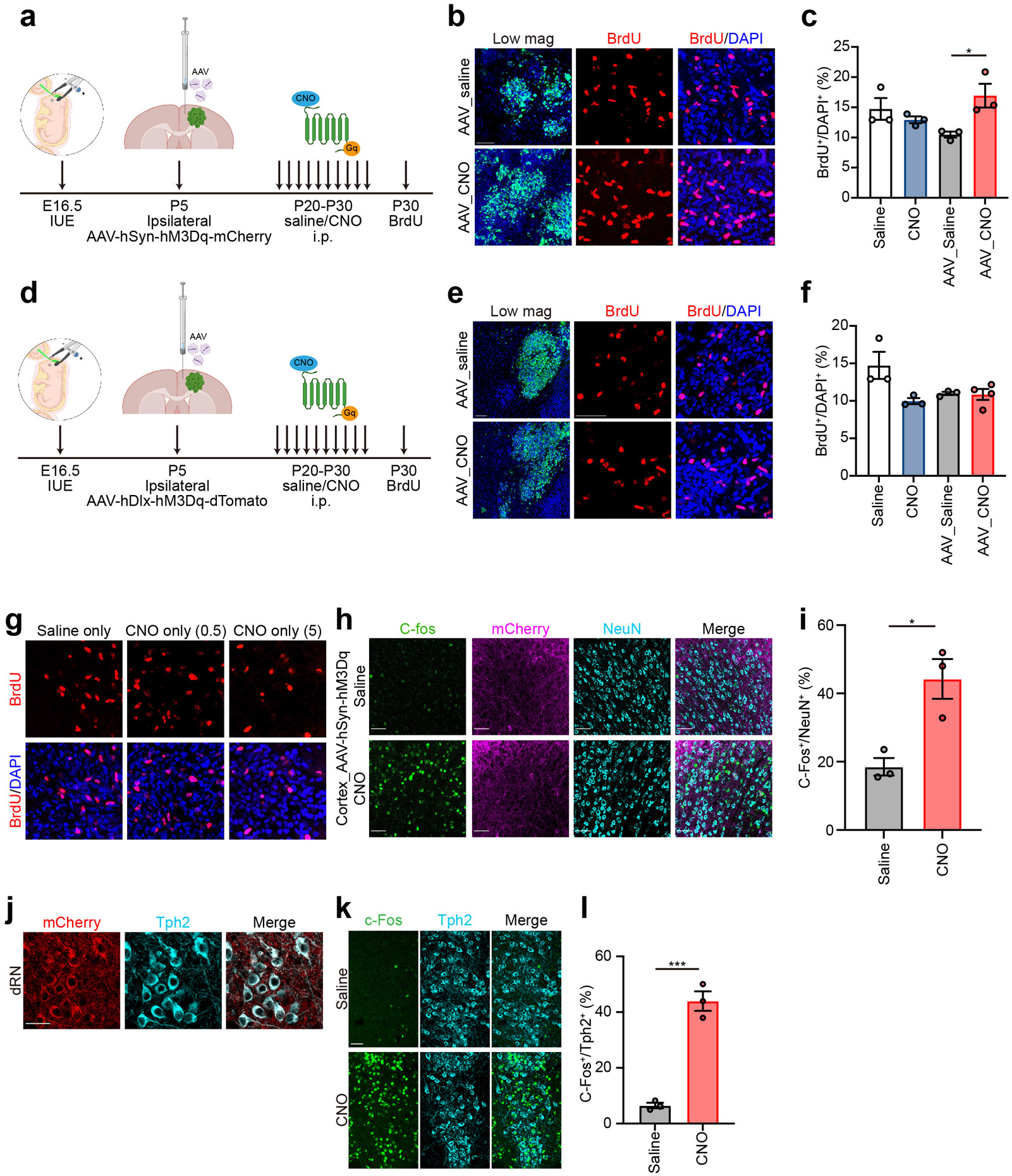
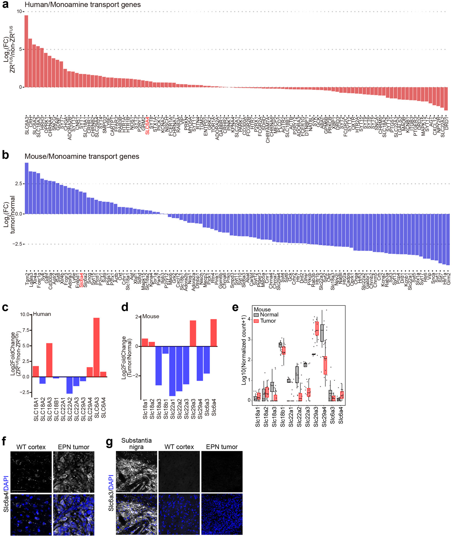
Bidirectional communication between tumours and neurons has emerged as a key facet of the tumour microenvironment that drives malignancy1,2. Another hallmark feature of cancer is epigenomic dysregulation, in which alterations in gene expression influence cell states and interactions with the tumour microenvironment3. Ependymoma (EPN) is a paediatric brain tumour that relies on epigenomic remodelling to engender malignancy4,5; however, how these epigenetic mechanisms intersect with extrinsic neuronal signalling during EPN tumour progression is unknown. Here we show that the activity of serotonergic neurons regulates EPN tumorigenesis, and that serotonin itself also serves as an activating modification on histones. We found that inhibiting histone serotonylation blocks EPN tumorigenesis and regulates the expression of a core set of developmental transcription factors. High-throughput, in vivo screening of these transcription factors revealed that ETV5 promotes EPN tumorigenesis and functions by enhancing repressive chromatin states. Neuropeptide Y (NPY) is one of the genes repressed by ETV5, and its overexpression suppresses EPN tumour progression and tumour-associated network hyperactivity through synaptic remodelling. Collectively, this study identifies histone serotonylation as a key driver of EPN tumorigenesis, and also reveals how neuronal signalling, neuro-epigenomics and developmental programs are intertwined to drive malignancy in brain cancer.


Figures

Extended Data Figure 1.
Extended Data Figure 1.. DREADD-based activation of neurons via CNO treatment
a. Schematic of DREADD-hM3Dq activation of ipsilateral cortical neurons in ZRFUS EPN mice. b. Low magnification image of EPN tumors and representative BrdU staining of EPN tumors after DREADD-hM3Dq activation of ipsilateral cortical neurons via CNO (scale bar=50 μm). c. Quantification of BrdU staining in saline versus CNO treated EPN tumors (n=3 per group, mean±SEM, unpaired two-sided Student’s t test, PSalinevsAAV_CNO = 0.4522, PCNOvsAAV_CNO = 0.1221, *PAAV_SalinevsAAV_CNO = 0.0337). d. Schematic of DREADD-hM3Dq activation of ipsilateral inhibitory neurons in ZRFUS EPN mice. e. Low magnification image of EPN tumors and representative BrdU staining of EPN tumors after DREADD-based activation of ipsilateral inhibitory neurons via CNO (scale bar=50 μm). f. Quantification of BrdU staining in saline versus CNO treated EPN tumors (Saline: n=3, CNO: n=3, AAV_Saline: n=3, AAV_CNO: n=4, mean±SEM, unpaired two-sided Student’s t test, PSalinevsAAV_CNO = 0.0770, PCNOvsAAV_CNO = 0.3896, PAAV_SalinevsAAV_CNO = 0.8727). g. Representative BrdU staining in saline and CNO (0.5 and 5 mg/kg) treated EPN tumors (scale bar=50 μm). h. Immunofluorescence staining of c-Fos in the ipsilateral cortical neurons in saline versus CNO treated EPN tumors (scale bar=50 μm). i. Quantification of c-Fos positive neurons in the ipsilateral cortex in saline versus CNO treated EPN tumors (n=3 per group, mean±SEM, unpaired two-sided Student’s t test, *P = 0.0155). j. Immunofluorescence staining of mCherry and Tph2 in the AAV injected dRN (scale bar=30 μm). Tph2: tryptophan hydroxylase 2. k. Immunofluorescence staining of c-Fos in the dRN serotonergic neurons in saline versus CNO treated EPN tumors (scale bar=50 μm). l. Quantification of c-Fos positive neurons in the dRN serotonergic neurons in saline versus CNO treated EPN tumors (n=3 per group, mean±SEM, unpaired two-sided Student’s t test, ***P = 0.0005). Panel a, d were created using Biorender.com
Extended Data Figure 2.
Extended Data Figure 2.. Expression of monoamine transporters in EPN
a. Expression levels of genes under monoamine transporter GO term in human ZRFUS versus non-ZRFUS EPN tumors. b. Expression levels of genes under monoamine transporter GO term in mouse EPN tumors versus non-tumor tissues. c. Log2(FC) of serotonin transporters in human ZRFUS tumors versus non-ZRFUS tumor tissues. d. Log2(FC) of serotonin transporters in mouse EPN tumors versus non-tumor tissues. e. Normalized read counts of serotonin transporters in mouse EPN tumors versus non-tumor tissues (at least n=10 per group, median±upper/lower limits, box boundary states upper/lower quartiles). f. Immunofluorescence staining of Slc6a4 in mouse non-tumor cortex and EPN tumors (scale bar=50 μm). g. Immunofluorescence staining of dopamine transporter (Slc6a3) in mouse substantia nigra, non-tumor cortex and EPN tumors (scale bar=50 μm).
Extended Data Figure 3.
Extended Data Figure 3.. Synaptic staining and H3-5HT staining after dRN manipulation
a. Low magnification image of tumor margin and representative higher magnification images (derived from dashed box) of peri-tumoral excitatory synaptic staining in saline versus CNO treated tumors from Fig. 1f (scale bar=10 μm). b. Quantification of excitatory synaptic staining in saline versus CNO treated tumors Fig. 1f (AAV_saline: n=4, AAV_CNO: n=3, mean±SEM, two-sided Wilcoxon rank sum test, P = 0.6286). c. Low magnification view of tumor margin and representative higher magnification images (derived from dashed box) of peri-tumoral inhibitory synaptic staining in saline versus CNO treated tumors from Fig. 1f (scale bar = 10 μm). d. Quantification of inhibitory synaptic staining in saline versus CNO treated tumors from Fig. 1f (AAV_saline: n=4, AAV_CNO: n=3, mean±SEM, unpaired two-sided Student’s t test, P = 0.7481). e. Low magnification image of tumor margin and representative higher magnification images (derived from dashed box) of peri-tumoral excitatory synaptic staining in saline versus CNO treated tumors from Fig. 1i (scale bar=10 μm). f. Quantification of excitatory synaptic staining in saline versus CNO treated tumors from Fig. 1i (n=3 per group, mean±SEM, unpaired two-sided Student’s t test, P = 0.6460). g. Low magnification image of tumor margin and representative higher magnification images (derived from dashed box) of peri-tumoral inhibitory synaptic staining in saline versus CNO treated tumors from Fig. 1i (scale bar=10 μm). h. Quantification of inhibitory synaptic staining in saline versus CNO treated tumors from Fig. 1i (n=3 per group, mean±SEM, unpaired two-sided Student’s t test, P = 0.9864). i. Immunofluorescence staining of H3-5HT in saline versus CNO treated tumors from Fig. 1f (scale bar=10 μm). j. Quantification of H3-5HT intensity in saline versus CNO treated tumors from Fig. 1f (n=3 per group, mean±SEM, unpaired two-sided Student’s t test, P = 0.2894). k. Immunofluorescence staining of H3-5HT in saline versus CNO treated tumors from Fig. 1i (scale bar=10 μm). l. Quantification of H3-5HT intensity in saline versus CNO treated tumors from Fig. 1i (n=3 per group, mean±SEM, unpaired two-sided Student’s t test, *P = 0.0465).
Extended Data Figure 4.
Extended Data Figure 4.. Tumor proliferation and Etv5 expression in H3.3-Q5A EPN tumors
a. Immunofluorescence staining of H3-5HT in H3.3-WT and H3.3-Q5A EPN tumors (scale bar=25 μm). b. Quantification of H3-5HT intensity in H3.3 control and H3.3-Q5A (n=4 per group, mean±SEM, two-sided Wilcoxon rank sum test, *P = 0.0286) c. Representative Ki67 staining of EPN control versus H3.3 wild-type and H3.3-Q5A tumors (scale bar=50 μm). d. Quantification of Ki67 staining in EPN control versus H3.3 wild-type and H3.3-Q5A tumors (EPN control: n=3, H3.3 wild-type: n=5, H3.3-Q5A: n=4, mean±SEM, unpaired two-sided Student’s t test, PEPNcontrolvsH3.3wild-type = 0.9040, **PEPNcontrolvsH3.3-Q5A = 0.0015, ****PH3.3wild-typevsH3.3-Q5A = 1.59E-05). e. Representative ETV5 staining of EPN control versus H3.3 wild-type and H3.3-Q5A tumors (scale bar=25 μm). f. Quantification of Etv5 staining in EPN control versus H3.3 wild-type and H3.3-Q5A tumors (EPN control: n=3, H3.3 wild-type: n=3, H3.3-Q5A: n=4, mean±SEM, unpaired two-sided Student’s t test, PEPNcontrolvsH3.3wild-type = 0.6180, **PEPNcontrolvsH3.3-Q5A = 0.0053, *PH3.3wild-typevsH3.3-Q5A = 0.0398).
Extended Data Figure 5.
Extended Data Figure 5.. Slc6a4-LOF EPN analyses, expression of serotonin synthetase, and SSRI treatment in EPN
a. Kaplan–Meier survival curve of EPN control (n=19, median=74 days) and Slc6a4-LOF (n=14, median=95 days, log-rank test, P = 0.1177). b. Representative Ki67 staining of EPN control versus Slc6a4-LOF tumors (scale bar=50 μm). c. Quantification of Ki67 staining in EPN control versus Slc6a4-LOF tumors (n=3 per group, mean±SEM, unpaired two-sided Student’s t test, **P = 0.0055). d. Representative H3-5HT staining of EPN control versus Slc6a4-LOF tumors (scale bar=25 μm). e. Quantification of H3-5HT intensity in EPN control versus Slc6a4-LOF tumors (n=4 per group, mean±SEM, two-sided Wilcoxon rank sum test, *P = 0.0286). f. Immunofluorescent staining of Tph2 in mouse dRN (positive control), non-tumor cortex, and EPN tumor (scale bar=50 μm) g. Quantification of Tph2 intensity in mouse dRN, non-tumor cortex, and EPN tumor (n=3 per group, mean±SEM, unpaired two-sided Student’s t test, ****PdRNvsnon-tumorcortex = 3.76E-05, **PdRNvsEPNtumor = 3.73E-05, Pnon-tumorcortexvsEPNtumor = 0.7262). h. Schematic of DMSO/SSRI treatment in EPN i. Representative H3-5HT staining of DMSO versus Sertraline-HCl treated tumors (scale bar=25 μm). j. Quantification of H3-5HT intensity in DMSOl versus Sertraline-HCl treated tumors (n=3 per group, mean±SEM, unpaired two-sided Student’s t test, P = 0.0743) k. Panel h was created using Biorender.com
Extended Data Figure 6.
Extended Data Figure 6.. H3-5HT ChIP-seq and ETV5 ChIP-seq analyses in EPN
a. Venn diagram depicting core TFs with H3-5HT peaks in mouse EPN tumors. b. ETV5 ChIP-seq heatmap profiles in mouse EPN tumors and venn diagram depicting genes overlapped between downregulated DEGs acquiring H3K27me3 peaks in ETV5-GOF tumors and ETV5 annotated genes.
Extended Data Figure 7.
Extended Data Figure 7.. Validation of candidates from functional screen
a. Immunoblots of Lhx2, Lhx4, Etv5, and Klf12 in mouse non-tumor cortex versus EPN tumors (n=3 per group). b. Kaplan–Meier survival curves of EPN control (n=51, median=70 days), LHX2-GOF (n=23, median=82 days, log-rank test, P = 0.9640), LHX4-GOF (n=9, median=114 days, log-rank test, P = 0.3532), and KLF12-GOF (n=23, median=77 days, log-rank test, P = 0.7588). c. Immunoblots of LHX2, LHX4, and KLF12 in control versus corresponding GOF tumors. d. Immunoblots of ETV5 in control versus ETV5-GOF and ETV5-LOF tumors. e. RT-qPCR fold-enrichment of ETV5 and Etv5 transcript (ddCt) in control versus ETV5-GOF and ETV5-LOF tumors (n=3 per group, mean±SEM, unpaired two-sided Student’s t test, GOF: **P = 0.0068, LOF: ***P = 0.0007). f. Surveyor Nuclease Digestion assay of Etv5 gRNA efficiency. Mouse non-tumor cortex: negative control. Two primer sets were used, and gel images are presented in left and right panel. Asterisks label the nuclease digested bands. g. Representative BrdU staining of control versus ETV5-GOF and ETV5-LOF tumors (scale bar=50 μm). h. Quantification of BrdU staining in control versus ETV5-GOF and ETV5-LOF tumors (n=3 per group, mean±SEM, unpaired two-sided Student’s t test, GOF: **P = 0.00258, LOF: ***P = 0.000976). i. Immunoblots of H3K27ac and H3K27me3 in control versus ETV5GOF tumors. Total H3: loading control. j. Ring chart showing percentage of H3K27me3 sites in ETV5-GOF tumors carrying ETS motif allowing 0 mismatch at 1,000 bp from peak center. k. GO-terms analysis of ETV5 binding partners in mouse EPN tumors performed using Enrichr (two-sided Fisher’s exact test). l. RT-qPCR fold-enrichment of gene transcript (ddCt) in control versus ETV5GOF tumors (n=3 per group, mean±SEM, unpaired two-sided Student’s t test, Npy: ***P = 0.00024, Gabra5: P = 0.4342, Chrm4: *P = 0.0239, Kcnmb4: *P = 0.0109, Nptx2: P = 0.2549). m. H3K27me3 ChIP-seq peaks at Npy and Chrm4 locus in control and ETV5-GOF tumors.
Extended Data Figure 8.
Extended Data Figure 8.. Kaplan–Meier survival curves comparison between sexes
a-c. All Kaplan–Meier survival curves, table of median survival, and log-rank test results comparison between groups divided by sex.
Extended Data Figure 9.
Extended Data Figure 9.. Analysis of NPY-GOF EPN tumors
a. RT-qPCR fold-enrichment of NPY transcript (ddCt) in control versus NPY-GOF tumors (n=3 per group, mean±SEM, unpaired two-sided Student’s t test, *P = 0.0255). b. RT-qPCR fold-enrichment of Npy2r transcript (ddCt) in mouse non-tumor cortex and EPN tumors (n=3 per group, as mean±SEM, unpaired two-sided Student’s t test, *P = 0.0215). c. Immunofluorescence staining of Npy2r in mouse cortex. NeuN: neuronal marker, Aldh1l1: astrocytic marker (scale bar=50 μm). d. Low magnification image of the tumor margin and representative images of Npy2r staining in mouse non-tumor and EPN tumors (scale bar=50 μm). Top right panel: Quantification of Npy2r intensity normalized to DAPI in mouse non-tumor cortex versus EPN tumors. (n=4 per group, mean±SEM, unpaired two-sided Student’s t test, **P = 0.0014). e. GO-terms analysis of the downregulated DEGs in NPY-GOF tumors versus control performed using Enrichr (two-sided Fisher’s exact test). f. Zoom-out EEG traces from mice bearing control and NPY-GOF tumors.
Figure 1.
Figure 1.. Stimulation of serotonergic neurons suppresses EPN tumorigenesis
a. GO-term analysis of up-regulated genes (log2(fold change(FC)) >=1, P < 0.05) in ZRFUS patients using GO Project datasets from Mouse Genome Informatics (MGI). b. Immunofluorescence staining of SLC6A4 in PFA and ZRFUS EPN patient samples (scale bar=25 μm). c. Schematic of DREADD-hM3Dq activation of ipsilateral excitatory neurons in ZRFUS EPN mice. d. Low magnification image of EPN tumors and representative BrdU staining of EPN tumors after DREADD-based activation of ipsilateral excitatory neurons via CNO (scale bar=50 μm). e. Quantification of BrdU staining in saline versus CNO treated EPN tumors (Saline: n=3, CNO: n=3, AAV_Saline: n=4, AAV_CNO: n=4, mean±SEM, unpaired Student’s two-sided t test, *PSalinevsAAV_CNO = 0.0289, *PCNOvsAAV_CNO = 0.0212, *PAAV_SalinevsAAV_CNO = 0.0132). f. Schematic of DREADD-hM3Dq activation of dRN neurons in ZRFUS EPN mice. g. Low magnification image of EPN tumors and representative BrdU staining of EPN tumors with DREADD-based activation of dRN neurons via CNO (scale bar=50 μm). h. Quantification of BrdU staining in saline versus CNO treated EPN tumors (Saline: n=3, CNO: n=3, AAV_saline: n=4, AAV_CNO: n=3, mean±SEM, unpaired Student’s t two-sided test, **PSalinevsAAV_CNO = 0.0024, ****PCNOvsAAV_CNO < 0.0001, ****PAAV_SalinevsAAV_CNO = 6.86E-05). i. Schematic of DREADD-hM4Di inhibition of dRN neurons in ZRFUS EPN mice. j. Low magnification image of EPN tumors and representative BrdU staining of EPN tumors with DREADD-based inhibition of dRN neurons via CNO (scale bar=50 μm). k. Quantification of BrdU staining in saline versus CNO treated EPN tumors (n=3 per group, mean±SEM, unpaired Student’s two-sided t test, **PSalinevsAAV_CNO = 0.01, **PCNOvsAAV_CNO = 0.0048, *PAAV_SalinevsAAV_CNO = 0.0133). Panel c, f, i were created using Biorender.com
Figure 2.
Figure 2.. Histone serotonylation is required for EPN tumorigenesis
a. Immunofluorescence staining of H3-5HT in PFA and ZRFUS EPN patient samples (scale bar=50 μm). b. Immunofluorescence staining of 5HT and H3-5HT in mouse EPN tumors (scale bar=25 μm). c. Immunoblots of histone serotonylation marks in mouse non-tumor cortex and EPN tumors (n=3 per group). d. Schematic of mouse EPN tumors expressing wild-type (H3.3) and dominant negative form (H3.3-Q5A) of histone variants. e. Representative low magnification image of H3.3 wild-type (P40) and H3.3-Q5A (P160) tumor (scale bar=50 μm), Kaplan–Meier survival curve of EPN H3.3 wild-type (n=10, median=80.5 days) and H3.3-Q5A (n=17, median=undefined, log-rank test, ***P = 0.0008), and table of tumor bearing mice vs all mice. f. ChIP-seq heatmap profiles demonstrating co-occupancy between ZRFUS-HA, H3-5HT, and H3K27ac in mouse EPN tumors. g. ZRFUS-HA, H3-5HT and H3K27ac ChIP-seq peaks at Ccnd1 locus. h. Significant TF motif (P < 0.05, cumulative binomial distribution test in HOMER software suite) of the genes annotated with ZRFUS-HA and H3-5HT peaks in mouse tumors. i. Venn diagram depicting core TFs annotated with H3-5HT peaks in mouse tumors and core TFs identified in ZRFUS patients from previous study. j. Representative core TF Etv5 locus with ZRFUS-HA, H3-5HT and H3K27ac ChIP-seq peaks. Panel k was created using Biorender.com
Figure 3.
Figure 3.. ETV5 regulates EPN progression and repressive chromatin states
a. Schematic of in vivo screening and barcode enrichment from mouse EPN tumors (n=5, unpaired two-sided Student’s t test). b. Kaplan–Meier survival curve of EPN control (n=51, median=70 days), ETV5-GOF (n=37, median=59 days, log-rank test, *P = 0.0167), and Etv5-LOF (n=40, median=92 days, log-rank test, *P = 0.0191). c. Comparison of H3K27ac and H3K27me3 ChIP-seq heatmap profiles between control versus ETV5-GOF tumors. TSS: transcription start site, TES: transcription end site. d. Schematic of ETV5 IP-MS in mouse non-tumor cortex and EPN tumors (n=3 per group). e. Volcano plot depicting ETV5 interactome in mouse EPN tumors (log2FC >=1, P < 0.05, two-sided Wald test, fold change compared to control samples). f. Venn diagram depicting ETV5 binding partners in mouse non-tumor cortex and EPN tumors. g. Left panel: Immunoblots of ZRFUS-HA, Etv5, Hdac1, and Cbx3 from mouse non-tumor cortex and EPN tumors. Right panel: Immunoprecipitation of Cbx3 and immunoblot of Etv5, Hdac1, and Cbx3 in mouse non-tumor cortex and EPN tumors. Arrowhead labels the protein of interest. h. Volcano plot of RNA-seq analysis from ETV5-GOF tumors versus control (n=3 per group, two-sided Wald test, log2FC >=1 or =< −1, P < 0.05). i. Venn diagram depicting downregulated DEGs acquiring H3K27me3 peaks in ETV5-GOF tumors. j. GO-terms analysis of the overlapping genes from i. performed using datasets from Enrichr (two-sided Fisher’s exact test). k. RT-qPCR fold-enrichment of NPY transcript (ddCT) in human normal brain and supratentorial EPN tissues (n=3 per group, mean±SEM, unpaired two-sided Student’s t test, ****P = 2.98E-06). l. Immunofluorescence staining of NPY in control versus ETV5-GOF tumors (scale bar=50 μm). m. Quantification of NPY in control versus ETV5-GOF tumors (n=4 per group, mean±SEM, unpaired two-sided Student’s t test. **P = 0.0031). Panel a, d were created using Biorender.com
Figure 4.
Figure 4.. NPY suppresses EPN progression
a. Kaplan–Meier survival curve of EPN control (n=11, median=70 days) and NPY-GOF (n=15, median=95 days, log-rank test, *P = 0.0174). b. Representative BrdU staining of control versus NPY-GOF tumors (scale bar=50 μm). c. Quantification of BrdU staining in control versus NPY-GOF tumors (n=3 per group, mean±SEM, unpaired two-sided Student’s t test, ***P = 0.0008). d. Volcano plot of RNA-seq analysis from NPY-GOF tumors versus control (n=3 per group, two-sided Wald test, log2FC >=1 or =< −1, P < 0.05). e. Low magnification view of tumor margin and representative higher magnification images (derived from dashed box) of peri-tumoral inhibitory synaptic staining in control versus NPY-GOF tumors (scale bar=25 μm). Vgat: vesicular GABA transporter. f. Quantification of inhibitory synaptic staining in control versus NPY-GOF tumors (n=5 per group, mean±SEM, two-sided Wilcoxon rank sum test, P = 0.0556). g. Low magnification image of the tumor margin and representative higher magnification images (derived from dashed box) of peri-tumoral excitatory synaptic staining in control versus NPY-GOF tumors (scale bar=25 μm). Vglut1: Vesicular glutamate transporter 1; Psd95: postsynaptic density protein 95. h. Quantification of excitatory synaptic staining in control versus NPY-GOF tumors (control: n=6, NPY-GOF: n=5, mean±SEM, two-sided Wilcoxon rank sum test, *P = 0.0173).
Figure 5.
Figure 5.. NPY suppresses EPN-induced brain hyperactivity
a. Schematic of electrophysiology recording in mCherry-labeled neurons around ZRFUS EPN tumor. b. Schematic of EEG recording in ZRFUS EPN mice. c. Traces of sEPSC recording in control and NPY-GOF mice d. Amplitude of sEPSC recording in control and NPY-GOF mice (n=4 mice per group, mean±SEM, unpaired two-sided Student’s t test, P = 0.8164). e. Frequency of sEPSC recording in control and NPY-GOF mice (n=4 mice per group, mean±SEM, two-sided Wilcoxon rank sum test, *P = 0.0286). f. Traces of sIPSC recording in control and NPY-GOF mice g. Amplitude of sIPSC recording in control and NPY-GOF mice (n=4 mice per group, mean±SEM, unpaired two-sided Student’s t test, P = 0.9893). h. Frequency of sIPSC recording in control and NPY-GOF mice (n=4 mice per group, mean±SEM, unpaired two-sided Student’s t test, *P = 0.0377). i. Seizure incidence curves across EEG recording sessions (control: n=8, NPY-GOF: n=6). j. Quantification of spikes number per hour over 48 hours period at 10-day intervals from P30 to P62 (data were derived from at least 3 mice in each group and are presented in violin plot with all individual data points, unpaired two-sided Student’s t test, P32: *P = 0.0290, P42: P = 0.7582, P52: P = 0.7903). k. Representative EEG traces from mice bearing control and NPY-GOF tumors. l. Model figure Panel a, b, l were created using Biorender.com

References

    1. Mancusi R & Monje M The neuroscience of cancer. Nature 618, 467–479 (2023). - PMC - PubMed
    1. Winkler F. et al. Cancer neuroscience: State of the field, emerging directions. Cell 186, 1689–1707 (2023). - PMC - PubMed
    1. Hanahan D. Hallmarks of Cancer: New Dimensions. Cancer Discov 12, 31–46 (2022). - PubMed
    1. Mack SC et al. Epigenomic alterations define lethal CIMP-positive ependymomas of infancy. Nature 506, 445–450 (2014). - PMC - PubMed
    1. Zhao S. et al. Epigenetic Alterations of Repeated Relapses in Patient-matched Childhood Ependymomas. Nat Commun 13, 6689 (2022). - PMC - PubMed

Methods References

    1. Yu G, Wang LG, Han Y & He QY clusterProfiler: an R package for comparing biological themes among gene clusters. OMICS 16, 284–287 (2012). - PMC - PubMed
    1. Sardar D. et al. Sox9 directs divergent epigenomic states in brain tumor subtypes. Proc Natl Acad Sci U S A 119, e2202015119 (2022). - PMC - PubMed
    1. Sengupta A & Holmes A A Discrete Dorsal Raphe to Basal Amygdala 5-HT Circuit Calibrates Aversive Memory. Neuron 103, 489–505 e487 (2019). - PMC - PubMed
    1. Langmead B & Salzberg SL Fast gapped-read alignment with Bowtie 2. Nat Methods 9, 357–359 (2012). - PMC - PubMed
    1. Li H. et al. The Sequence Alignment/Map format and SAMtools. Bioinformatics 25, 2078–2079 (2009). - PMC - PubMed

MeSH terms