

Inhibition of insulin degrading enzyme suppresses osteoclast hyperactivity via enhancing Nrf2-dependent antioxidant response in glucocorticoid-induced osteonecrosis of the femoral head
- PMID: 39085816
- PMCID: PMC11292917
- DOI: 10.1186/s10020-024-00880-1
Inhibition of insulin degrading enzyme suppresses osteoclast hyperactivity via enhancing Nrf2-dependent antioxidant response in glucocorticoid-induced osteonecrosis of the femoral head
Abstract
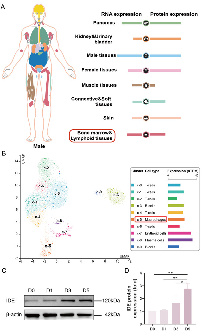
Background: Osteoclast hyperactivation due to the pathological overproduction of reactive oxygen species (ROS) stimulated by glucocorticoids (GCs) is one of the key drivers behind glucocorticoid-induced osteonecrosis of the femoral head (GIONFH). The insulin degrading enzyme (IDE), a conserved Zn2+ metallo-endopeptidase, facilitates the DNA binding of glucocorticoid receptor and plays a substantial role in steroid hormone-related signaling pathways. However, the potential role of IDE in the pathogenesis of GIONFH is yet undefined.
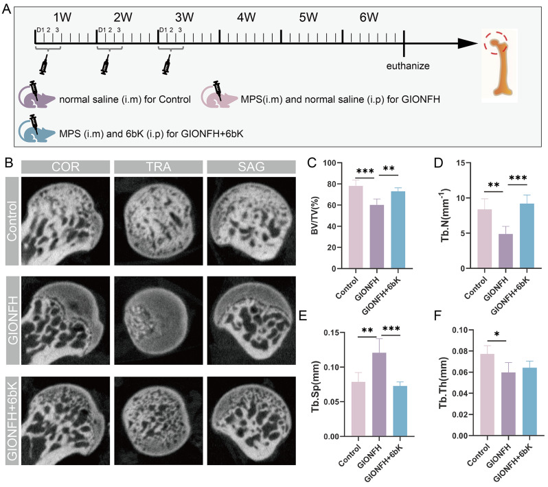
Methods: In this study, we employed network pharmacology and bioinformatics analysis to explore the impact of IDE inhibition on GIONFH with 6bK as an inhibitory agent. Further evidence was collected through in vitro osteoclastogenesis experiments and in vivo evaluations involving methylprednisolone (MPS)-induced GIONFH mouse model.
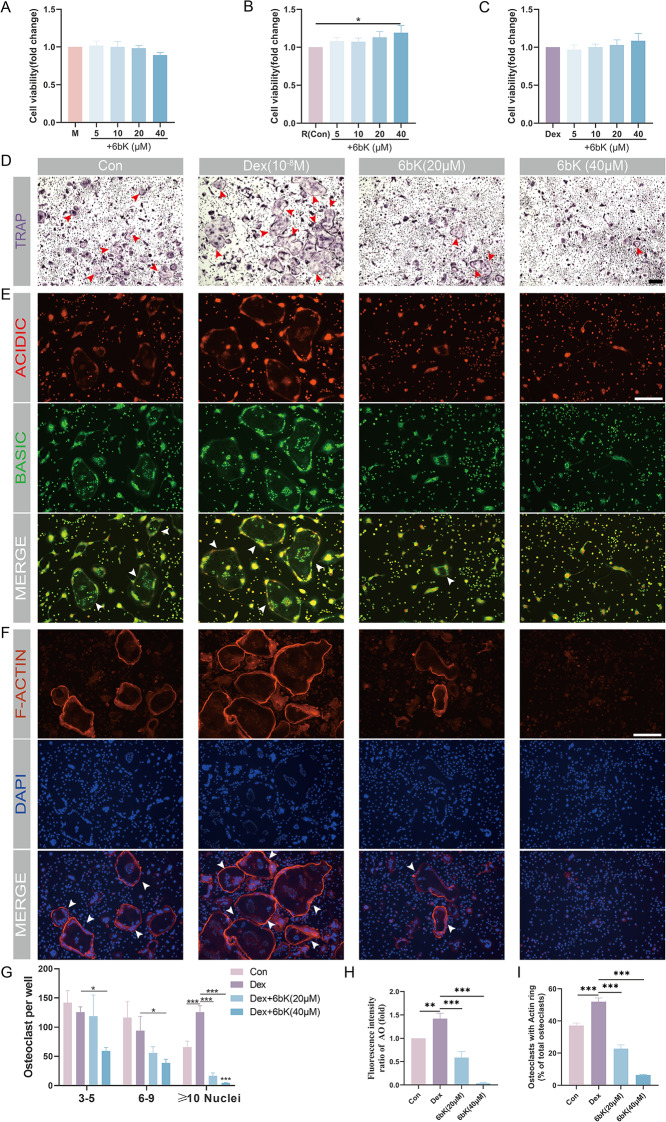
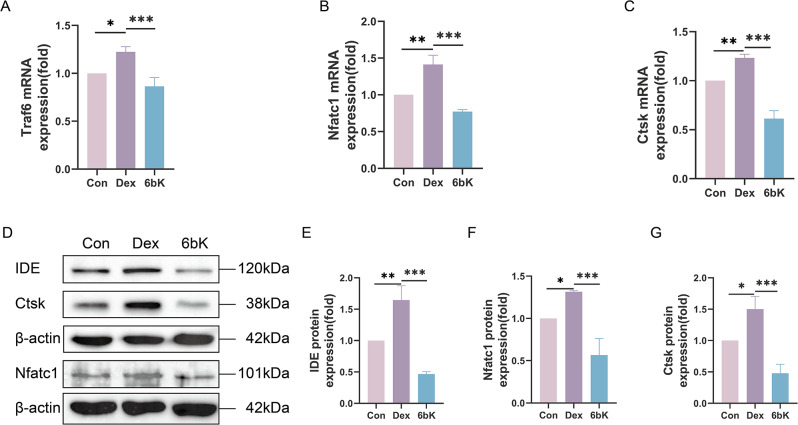
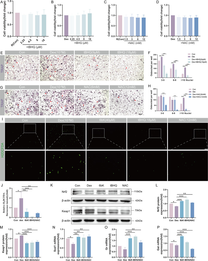
Results: Enrichment analysis indicated a potential role of 6bK in redox regulation amid GIONFH development. In vitro findings revealed that 6bK could attenuate GCs-stimulated overactivation of osteoclast differentiation by interfering with the transcription and expression of key osteoclastic genes (Traf6, Nfatc1, and Ctsk). The use of an H2DCFDA probe and subsequent WB assays introduced the inhibitory effects of 6bK on osteoclastogenesis, linked with the activation of the nuclear factor erythroid-derived 2-like 2 (Nrf2)-mediated antioxidant system. Furthermore, Micro-CT scans validated that 6bK could alleviate GIONFH in MPS-induced mouse models.
Conclusions: Our findings suggest that 6bK suppresses osteoclast hyperactivity in GCs-rich environment. This is achieved by reducing the accumulation of intracellular ROS via promoting the Nrf2-mediated antioxidant system, thus implying that IDE could be a promising therapeutic target for GIONFH.
Keywords: Glucocorticoid-induced osteonecrosis of the femoral head (GIONFH); Glucocorticoids (GCs); Insulin degrading enzyme (IDE); Osteoclast; Reactive oxygen species (ROS).
© 2024. The Author(s).
Conflict of interest statement
The authors declare that they have no competing interests.
Figures


Similar articles
-
Astaxanthin-mediated Nrf2 activation ameliorates glucocorticoid-induced oxidative stress and mitochondrial dysfunction and impaired bone formation of glucocorticoid-induced osteonecrosis of the femoral head in rats.J Orthop Surg Res. 2024 May 14;19(1):294. doi: 10.1186/s13018-024-04775-z. J Orthop Surg Res. 2024. PMID: 38745231 Free PMC article.
-
Inhibition of protein disulfide isomerase mitigates steroid-induced osteonecrosis of the femoral head by suppressing osteoclast activity through the reduction of cellular oxidative stress.Chem Biol Interact. 2024 Dec 1;404:111263. doi: 10.1016/j.cbi.2024.111263. Epub 2024 Oct 10. Chem Biol Interact. 2024. PMID: 39393751
-
Taxifolin-mediated Nrf2 activation ameliorates oxidative stress and apoptosis for the treatment of glucocorticoid-induced osteonecrosis of the femoral head.Phytother Res. 2024 Jan;38(1):156-173. doi: 10.1002/ptr.8031. Epub 2023 Oct 17. Phytother Res. 2024. PMID: 37846877
-
Naringin regulates bone metabolism in glucocorticoid-induced osteonecrosis of the femoral head via the Akt/Bad signal cascades.Chem Biol Interact. 2019 May 1;304:97-105. doi: 10.1016/j.cbi.2019.03.008. Epub 2019 Mar 14. Chem Biol Interact. 2019. PMID: 30878453
-
Vasculature deprivation--induced osteonecrosis of the rat femoral head as a model for therapeutic trials.Theor Biol Med Model. 2005 Jul 5;2:24. doi: 10.1186/1742-4682-2-24. Theor Biol Med Model. 2005. PMID: 15996271 Free PMC article. Review.
Cited by
-
Skimmianine Attenuates Osteoclast Activity by Suppressing ERp57-Driven Calcium Oscillations/Calcineurin/Nfatc1 Signalling in Postmenopausal Osteoporosis.J Cell Mol Med. 2025 Aug;29(15):e70777. doi: 10.1111/jcmm.70777. J Cell Mol Med. 2025. PMID: 40797290 Free PMC article.
-
Association between composite dietary antioxidant index and rheumatoid arthritis: results from NHANES 2003-2018.Int J Med Sci. 2025 Feb 18;22(5):1184-1193. doi: 10.7150/ijms.107332. eCollection 2025. Int J Med Sci. 2025. PMID: 40027194 Free PMC article.
-
Quercetagetin alleviates inflammatory osteoclastogenesis and collagen antibody-induced arthritis via Nrf2 signaling and Pten/AKT/Nfatc1 axis.Arthritis Res Ther. 2025 Mar 8;27(1):54. doi: 10.1186/s13075-025-03522-x. Arthritis Res Ther. 2025. PMID: 40057805 Free PMC article.
-
Decreased expression of insulin-degrading enzyme increases gluconeogenesis and glucose production in cultured hepatocytes administered with glucagon.Sci Rep. 2025 May 31;15(1):19168. doi: 10.1038/s41598-025-03790-2. Sci Rep. 2025. PMID: 40450129 Free PMC article.
References
-
- Baek JM, Kim JY, Yoon KH, Oh J, Lee MS. Ebselen is a potential Anti-osteoporosis Agent by suppressing receptor activator of nuclear factor Kappa-B ligand-Induced Osteoclast differentiation in vitro and Lipopolysaccharide-Induced Inflammatory Bone Destruction in vivo. Int J Biol Sci. 2016;12(5):478–88. 10.7150/ijbs.13815 - DOI - PMC - PubMed
-
- Chen J, Cui Z, Wang Y, Lyu L, Feng C, Feng D, et al. Cyclic polypeptide D7 protects bone marrow mesenchymal cells and promotes chondrogenesis during osteonecrosis of the femoral head via growth differentiation factor 15-Mediated Redox Signaling. Oxid Med Cell Longev. 2022;2022:3182368. - PMC - PubMed
MeSH terms
Substances
Grants and funding
LinkOut - more resources
Full Text Sources
Medical
Miscellaneous
