. 2024 Jul 31;23(1):151.
doi: 10.1186/s12943-024-02052-5.

Circular RNA hsa_circ_0000467 promotes colorectal cancer progression by promoting eIF4A3-mediated c-Myc translation

Affiliations

Circular RNA hsa_circ_0000467 promotes colorectal cancer progression by promoting eIF4A3-mediated c-Myc translation

Xianjie Jiang et al. Mol Cancer. .

Abstract

Background: Colorectal cancer (CRC) is the second most common malignant tumor worldwide, and its incidence rate increases annually. Early diagnosis and treatment are crucial for improving the prognosis of patients with colorectal cancer. Circular RNAs are noncoding RNAs with a closed-loop structure that play a significant role in tumor development. However, the role of circular RNAs in CRC is poorly understood.

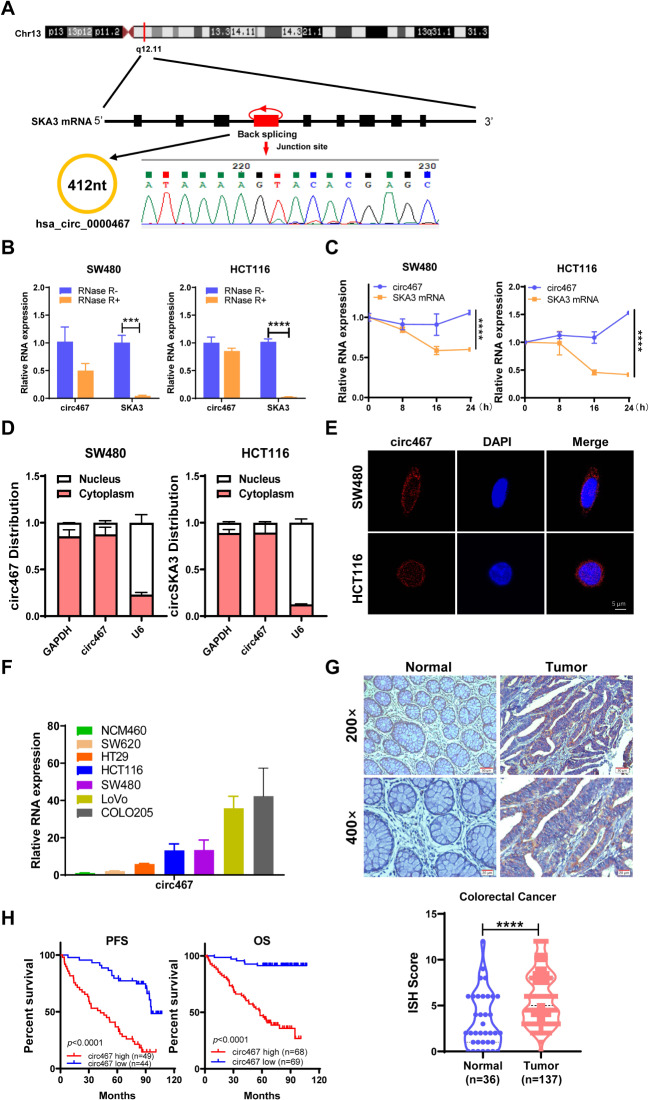
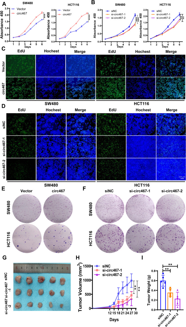
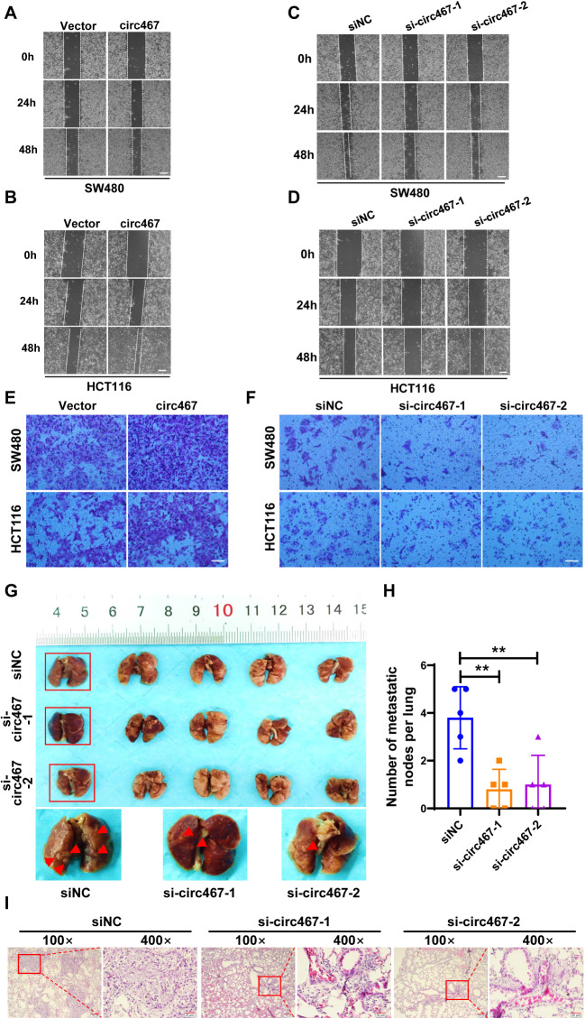
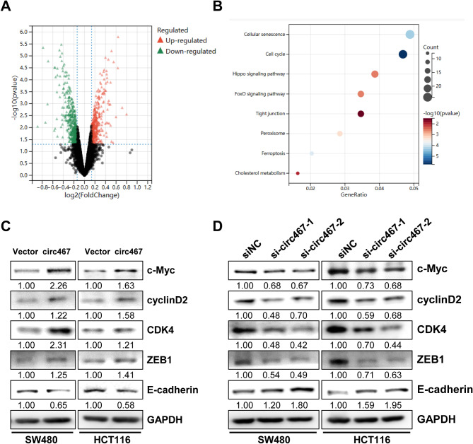
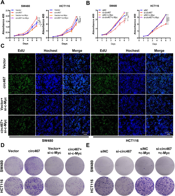
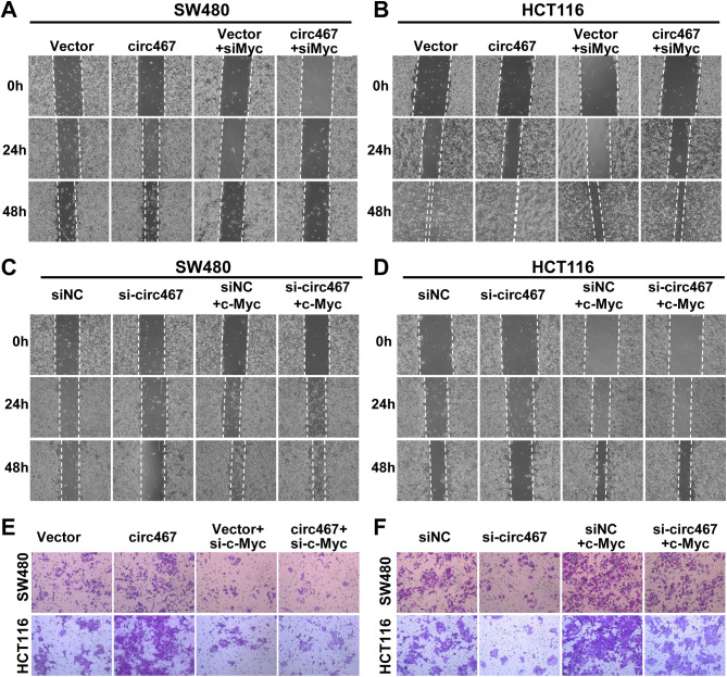
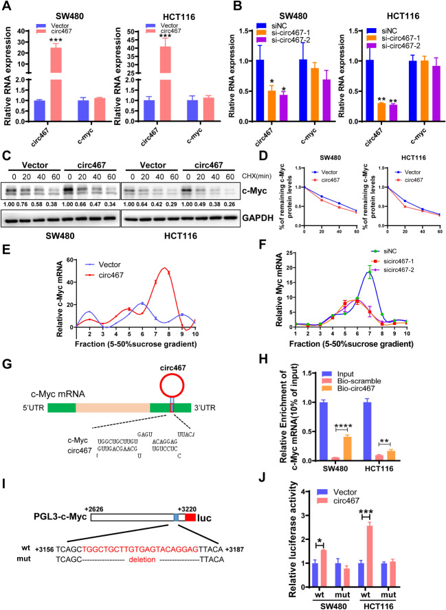
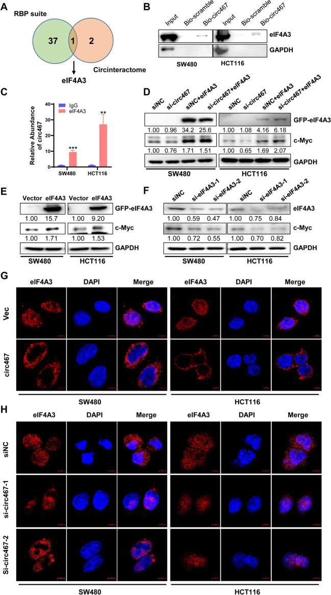
Methods: The circular RNA hsa_circ_0000467 was screened in CRC circRNA microarrays using a bioinformatics analysis, and the expression of hsa_circ_0000467 in CRC tissues was determined by in situ hybridization. The associations between the expression level of hsa_circ_0000467 and the clinical characteristics of CRC patients were evaluated. Then, the role of hsa_circ_0000467 in CRC growth and metastasis was assessed by CCK8 assay, EdU assay, plate colony formation assay, wound healing assay, and Transwell assay in vitro and in a mouse model of CRC in vivo. Proteomic analysis and western blotting were performed to investigate the effect of hsa_circ_0000467 on c-Myc signaling. Polysome profiling, RT‒qPCR and dual-luciferase reporter assays were performed to determine the effect of hsa_circ_0000467 on c-Myc translation. RNA pull-down, RNA immunoprecipitation (RIP) and immunofluorescence staining were performed to assess the effect of hsa_circ_0000467 on eIF4A3 distribution.

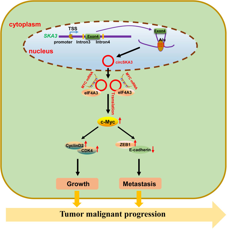
Results: In this study, we found that the circular RNA hsa_circ_0000467 is highly expressed in colorectal cancer and is significantly correlated with poor prognosis in CRC patients. In vitro and in vivo experiments revealed that hsa_circ_0000467 promotes the growth and metastasis of colorectal cancer cells. Mechanistically, hsa_circ_0000467 binds eIF4A3 to suppress its nuclear translocation. In addition, it can also act as a scaffold molecule that binds eIF4A3 and c-Myc mRNA to form complexes in the cytoplasm, thereby promoting the translation of c-Myc. In turn, c-Myc upregulates its downstream targets, including the cell cycle-related factors cyclin D2 and CDK4 and the tight junction-related factor ZEB1, and downregulates E-cadherin, which ultimately promotes the growth and metastasis of CRC.

Conclusions: Our findings revealed that hsa_circRNA_0000467 plays a role in the progression of CRC by promoting eIF4A3-mediated c-Myc translation. This study provides a theoretical basis and molecular target for the diagnosis and treatment of CRC.

Keywords: Colorectal cancer; Growth; Metastasis; Translation; c-Myc; eIF4A3; hsa_circRNA_0000467.


Conflict of interest statement

The authors declare that they have no competing interests.

Figures

Fig. 1
Fig. 1
Circ467 is highly expressed in CRC and is inversely correlated with the prognosis in CRC patients. (A) Schematic diagram of circ467 generation. (B) SW480 and HCT116 cells were treated with RNase R, and the stability of circ467 was assessed using RT‒qPCR. (C) SW480 and HCT116 cells were treated with actinomycin D for the indicated times, and the stability of circ467 was assessed using RT‒qPCR. (D) The cellular localization of circ467 was assessed by RNA nucleoplasmic separation assays. (E) The intracellular distribution of circ467 was assessed by fluorescence in situ hybridization (scale bar = 5 μm). (F) circ467 expression in CRC cells and NCM460 cells (immortalized colorectal epithelial cells) was assessed using RT‒qPCR. (G) Representative results showing circ467 expression in 137 CRC tissues and 36 adjacent nontumor tissues according to in situ hybridization (upper panel); circ467 was highly expressed in CRC tissues (lower panel). Magnification: 200×, scale bar = 50 μm; magnification: 400×; scale bar = 20 μm. (H) Kaplan‒Meier analysis of overall survival and progression-free survival of CRC patients grouped according to circ467 expression. The data shown are representative images or are expressed as the mean ± SD of each group from three separate experiments or a single experiment (for the in vivo studies) (***, p < 0.001; ****, p < 0.0001 vs. control; Student’s t test)
Fig. 2
Fig. 2
Circ467 promotes the growth of CRC cells in vitro and in vivo A, B. The proliferation of SW480 and HCT116 cells after circ467 overexpression or knockdown was assessed by CCK-8 assay. C, D. The proliferation of SW480 and HCT116 cells after circ467 overexpression or knockdown was assessed by EdU assay. E, F. Colony formation assays were performed in SW480 and HCT116 cells after circ467 overexpression or knockdown. G. Images of tumors formed in nude mice after transplantation of HCT116 cells transfected with circ467 siRNAs. H. Statistical analysis of the growth curves of tumors formed in nude mice after transplantation of HCT116 cells transfected with circ467 siRNAs. I. Statistical analysis of the weight of xenograft tissues in nude mice after transplantation of HCT116 cells transfected with circ467 siRNAs. The data shown are representative images or are expressed as the mean ± SD of each group from three experiments or a single experiment (for the in vivo studies) (**, p < 0.01; ***, p < 0.001; ****, p < 0.0001 vs. control; Student’s t test)
Fig. 3
Fig. 3
Circ467 promotes the metastasis of CRC in vitro and in vivo A, B. The migration of SW480 and HCT116 cells after circ467 overexpression was assessed using a wound healing assay. Scale bar = 200 μm. C, D. The migration of SW480 and HCT116 cells after circ467 knockdown was assessed using a wound healing assay. Scale bar = 200 μm. E, F. The invasiveness of SW480 and HCT116 cells after circ467 overexpression or knockdown was assessed by Transwell assay. Scale bar = 50 μm. G. Images of visible nodules on the lung surface after HCT116 cells in which circ467 was knocked down were injected into the tail vein of nude mice (n = 5); arrows show visible nodules on the lung surface. H. Quantification of metastatic nodules on the lung surface. I. Representative images of H&E-stained metastatic lung nodules after circ467 knockdown. Magnification = 100×, scale bar = 100 μm; magnification = 400×; scale bar = 20 μm. The data shown are representative images or are expressed as the mean ± SD of each group from a single experiment (for the in vivo studies) (**, p < 0.01 vs. control; Student’s t test)
Fig. 4
Fig. 4
Circ467 can activate c-Myc signaling (A) Volcano plot showing differentially expressed proteins in control and circ467-overexpressing HCT116 cells. (B) The principal biological processes regulated by circ467 were determined through KEGG enrichment analysis of the differentially expressed proteins between control and circ467-overexpressing HCT116 cells. (C) The expression of c-Myc, cyclin D2, CDK4, ZEB1, and E-cadherin in SW480 and HCT116 cells after circ467 overexpression was assessed by western blotting; GAPDH served as an internal control. (D) The expression of c-Myc, cyclin D2, CDK4, ZEB1, and E-cadherin in SW480 and HCT116 cells after circ467 knockdown was assessed by western blotting; GAPDH served as an internal control
Fig. 5
Fig. 5
Circ467 promotes the growth of CRC cells by upregulating c-Myc. (A) CCK-8 assays were performed to assess the growth of SW480 and HCT116 cells after cotransfection of circ467 plasmids and c-Myc siRNAs. (B) CCK-8 assays were performed to assess the growth of SW480 and HCT116 cells after cotransfection of circ467 siRNAs and c-Myc plasmids. (C) EdU assays were performed to assess the growth of SW480 and HCT116 cells after cotransfection of circ467 plasmids and c-Myc siRNAs. (D) Transwell assays were performed to assess the growth of SW480 and HCT116 cells after cotransfection of circ467 plasmids and c-Myc siRNAs. (E) Transwell assays were performed to assess the growth of SW480 and HCT116 cells after cotransfection of circ467 siRNAs and c-Myc plasmids. The data shown are representative images or are expressed as the mean ± SD of each group from three separate experiments (*, p < 0.05; **, p < 0.01; ***, p < 0.001; ****, p < 0.0001 vs. control; Student’s t test)
Fig. 6
Fig. 6
Circ467 promotes the migration and invasion of CRC cells by upregulating c-Myc. A, B. Wound healing assays were performed using SW480 and HCT116 cells after cotransfection with circ467 plasmids and c-Myc siRNAs. C, D. Wound healing assays were performed using SW480 and HCT116 cells after cotransfection with circ467 siRNAs and c-Myc plasmids. E. Transwell assays were performed using SW480 and HCT116 cells after cotransfection with circ467 plasmids and c-Myc siRNAs. F. Transwell assays were performed using SW480 and HCT116 cells after cotransfection with circ467 siRNAs and c-Myc plasmids. The data shown are representative images of three separate experiments from each group
Fig. 7
Fig. 7
Circ467 may increase the translation efficiency of c-Myc. A The expression of circ467 and c-Myc in SW480 and HCT116 cells after circ467 overexpression was assessed using RT‒qPCR. B The expression of circ467 and c-Myc in SW480 and HCT116 cells after circ467 knockdown was assessed using RT‒qPCR. C The expression of c-Myc in SW480 and HCT116 cells after circ467 overexpression and cycloheximide (CHX) treatment was assessed by western blotting. D Quantification of c-Myc protein degradation according to C. E, F. Polysomes in cytoplasmic extracts from HCT116 cells after circ467 overexpression or knockdown were fractionated by sucrose gradients, and the relative c-Myc mRNA expression level in the gradient fractions was analyzed using RT‒qPCR. G Diagram showing the interaction between circ467 and c-Myc mRNA. H The interaction between circ467 and c-Myc mRNA in SW480 and HCT116 cells was evaluated by RNA pull-down using an anti-circ467 probe, followed by RT‒qPCR analysis of c-Myc expression. I Schematic diagram of the construction of the wild-type and mutant c-Myc 3’ UTR reporters. J c-Myc reporter activity in SW480 and HCT116 cells cotransfected with the circ467 plasmid and potential c-Myc reporters (PGL3-c-Myc-wt or PGL3-c-Myc-mut) was assessed using a dual luciferase reporter assay. The data shown are representative images or are expressed as the mean ± SD of each group from three separate experiments (*, p < 0.05; **, p < 0.01; ***, p < 0.001; ****, p < 0.0001 vs. control, Student’s t test)
Fig. 8
Fig. 8
Circ467 upregulates c-Myc by binding to eIF4A3 and inhibiting its nuclear translocation A Venn diagram indicating that eIF4A3 is an RNA-binding protein of circ467 according to both RNA-binding protein databases. B The interaction between circ467 and eIF4A3 in SW480 and HCT116 cells was evaluated by RNA pull-down using an anti-circ467 probe, followed by western blotting analysis of eIF4A3. C The interaction between eIF4A3 and circ467 in SW480 and HCT116 cells was evaluated by RNA immunoprecipitation using an anti-eIF4A3 antibody, followed by RT‒qPCR. D The expression of GFP-eIF4A3 and c-Myc in SW480 and HCT116 cells after cotransfection of circ467 siRNAs and GFP-eIF4A3 plasmids was evaluated by western blotting. E, F The expression of eIF4A3 and c-Myc in SW480 and HCT116 cells after GFP-eIF4A3 overexpression or eIF4A3 knockdown was assessed by western blotting. G, H. The distribution of eIF4A3 in SW480 and HCT116 cells after circ467 overexpression or knockdown was assessed by immunofluorescence staining. The data shown are representative images or are expressed as the mean ± SD of each group from three separate experiments (**, p < 0.01; ***, p < 0.001 vs. control; Student’s t test)
Fig. 9
Fig. 9
Schematic representation of the possible mechanism of circ467 in the malignant progression of CRC

Similar articles

Cited by

References

    1. Sedlak JC, Yilmaz OH, Roper J. Metabolism and colorectal Cancer. Annu Rev Pathol. 2023;18:467–92. 10.1146/annurev-pathmechdis-031521-041113 - DOI - PMC - PubMed
    1. Sung H, Ferlay J, Siegel RL, Laversanne M, Soerjomataram I, Jemal A, et al. Global Cancer statistics 2020: GLOBOCAN estimates of incidence and Mortality Worldwide for 36 cancers in 185 countries. CA Cancer J Clin. 2021;71(3):209–49. 10.3322/caac.21660 - DOI - PubMed
    1. Arnold M, Sierra MS, Laversanne M, Soerjomataram I, Jemal A, Bray F. Global patterns and trends in colorectal cancer incidence and mortality. Gut. 2017;66(4):683–91. 10.1136/gutjnl-2015-310912 - DOI - PubMed
    1. Biller LH, Schrag D. Diagnosis and treatment of metastatic colorectal Cancer: a review. JAMA. 2021;325(7):669–85. 10.1001/jama.2021.0106 - DOI - PubMed
    1. Ciardiello F, Ciardiello D, Martini G, Napolitano S, Tabernero J, Cervantes A. Clinical management of metastatic colorectal cancer in the era of precision medicine. CA Cancer J Clin. 2022;72(4):372–401. 10.3322/caac.21728 - DOI - PubMed

MeSH terms

LinkOut - more resources