. 2024 Jun:30:101147.
doi: 10.1016/j.cossms.2024.101147. Epub 2024 Mar 18.

Advancements in fluorescence lifetime imaging microscopy Instrumentation: Towards high speed and 3D

Affiliations

Advancements in fluorescence lifetime imaging microscopy Instrumentation: Towards high speed and 3D

Jongchan Park et al. Curr Opin Solid State Mater Sci. 2024 Jun.

Abstract

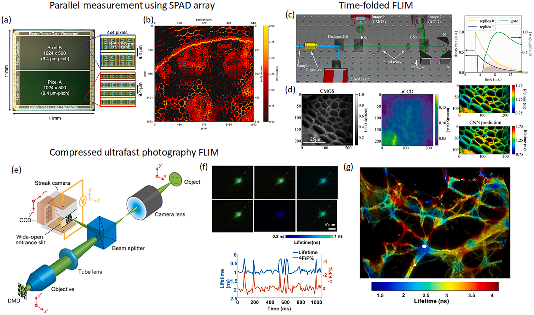
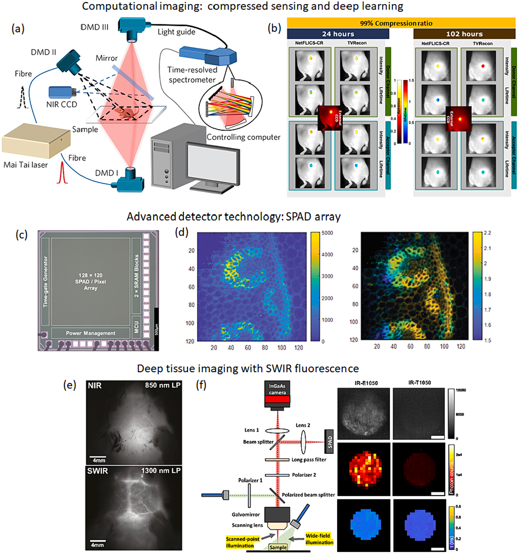
Fluorescence lifetime imaging microscopy (FLIM) is a powerful imaging tool offering molecular specific insights into samples through the measurement of fluorescence decay time, with promising applications in diverse research fields. However, to acquire two-dimensional lifetime images, conventional FLIM relies on extensive scanning in both the spatial and temporal domain, resulting in much slower acquisition rates compared to intensity-based approaches. This problem is further magnified in three-dimensional imaging, as it necessitates additional scanning along the depth axis. Recent advancements have aimed to enhance the speed and three-dimensional imaging capabilities of FLIM. This review explores the progress made in addressing these challenges and discusses potential directions for future developments in FLIM instrumentation.

Keywords: 3D imaging; Bioimaging; Fluorescence lifetime; High speed imaging; Microscopy.


Conflict of interest statement

Declaration of competing interest The authors declare that they have no known competing financial interests or personal relationships that could have appeared to influence the work reported in this paper.

Figures

Fig. 1.
Fig. 1.. Fluorescence lifetime imaging.
(a) FLIM records time-resolved fluorescence decay at each spatial location. (b) Frequency-domain FLIM. (c) Time-domain FLIM.
Fig. 2.
Fig. 2.
High-speed FLIM methods. (a) The photon rate (throughput) of TCSPC-mode FLIM is limited to avoid measurement errors such as pile-up effects and counting loss. (b) The photon rate can be increased by using TCSPC with parallel detector arrays [22]. (c) Fluorescence decay waveform can be directly measured by high-speed photodetectors [23]. (d) Two-photon fluorescence lifetime dynamics of apoptosis induced cells were recorded at a video rate [24]. (e) Spectro-temporal encoded illumination enables kilohertz FLIM [25] and its application in (f) imaging flow cytometry.
Fig. 3.
Fig. 3.
High-speed FLIM methods using 2D detector arrays. (a) 0.5-megapixel SPAD camera with an acquisition rate up to 1 Hz [28] (b) 3.6 megapixel wide-field FLIM image captured by using the SPAD camera with image stitching [27]. (c) Time-folded FLIM. An optical cavity generates temporally delayed and spatially sheared replicas of fluorescent decay signal [29]. (d) The signal from the CMOS sensor and intensified CCD was used to generate fluorescence lifetime image with either an inverse retrieval method or neural network [29]. (e) Compressed ultrafast photography FLIM. The fluorescence signal is spatially mapped by the DMD, temporally sheared by the streak camera, and mapped onto the CCD sensor [31]. The fluorescence lifetime image is computationally reconstructed by solving the inverse problem. (f) High-speed fluorescence lifetime imaging of neural action potentials in live cells [30]. (g) Fluorescence lifetime imaging of neuronal cytoskeleton immunolabeled with multiple fluorophores.
Fig. 4.
Fig. 4.
3D FLIM. (a) Light-sheet microscopy with a TCSPC-based wide-field FLIM detector [42]. (b) 3D volumetric fluorescence lifetime image of a mixture of fluorescent beads and quantum dots [42]. (c) 3D fluorescence lifetime imaging with optical projection tomography. A series of time-gated fluorescence images were acquired at various projection angles by rotating the sample [46]. (d) 3D volumetric fluorescence lifetime image of a live zebrafish [48]. (e) Light-field tomography FLIM. The 2D perspective images of a 3D sample are captured by pupil selection through a scanning mirror. The resultant images are further rotated and compressed along one spatial axis by a dove prism and cylindrical lens, respectively. (f) High-resolution depth-sectioned fluorescence lifetime images of a mouse kidney tissue section.
Fig. 5.
Fig. 5.
Future perspective. (a) Compressed sensing method to overcome the information limit in FLIM [60]. (b) In vivo FLIM with a data compression ratio of 99% [59]. (c) 3D-stacked SPAD image sensor with a 45% fill factor [63]. (d) Fluorescence lifetime imaging of Convallaria Majalis sample at a video rate using the SPAD array [63]. (e) The mouse brain vasculature imaging through skin and skull. SWIR fluorescence enables deep tissue imaging beyond the NIR window [69]. (f) Lifetime characterization of SWIR fluorophores [70].

References

    1. Berezin MY, Achilefu S, Fluorescence lifetime measurements and biological imaging, Chem. Rev 110 (2010) 2641–2684. - PMC - PubMed
    1. Datta R, Heaster TM, Sharick JT, Gillette AA, Skala MC, Fluorescence lifetime imaging microscopy: fundamentals and advances in instrumentation, analysis, and applications, J. Biomed. Opt 25 (2020) 071203. - PMC - PubMed
    1. Alfonso-Garcia A, Bec J, Weyers B, Marsden M, Zhou X, Li C, Marcu L, Mesoscopic fluorescence lifetime imaging: fundamental principles, clinical applications and future directions, J. Biophotonics 14 (2021) e202000472. - PMC - PubMed
    1. Dmitriev RI, Intes X, Barroso MM, Luminescence lifetime imaging of three-dimensional biological objects, J. Cell Sci 134 (2021) 1–17. - PMC - PubMed
    1. Le Marois A, Suhling K, Quantitative live cell FLIM imaging in three dimensions, Multi-Parametric Live Cell Microscopy of 3D Tissue Models (2017) 31–48. - PubMed

LinkOut - more resources