Review
. 2023 Aug 23:3:1235188.
doi: 10.3389/fmmed.2023.1235188. eCollection 2023.

Mitochondrial calcium signaling and redox homeostasis in cardiac health and disease

Affiliations
Review

Mitochondrial calcium signaling and redox homeostasis in cardiac health and disease

Tudor-Alexandru Popoiu et al. Front Mol Med. .

Abstract

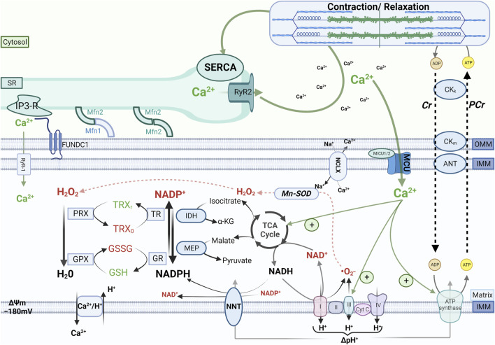
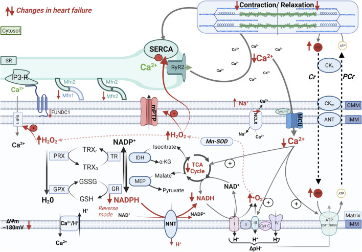
The energy demand of cardiomyocytes changes continuously in response to variations in cardiac workload. Cardiac excitation-contraction coupling is fueled primarily by adenosine triphosphate (ATP) production by oxidative phosphorylation in mitochondria. The rate of mitochondrial oxidative metabolism is matched to the rate of ATP consumption in the cytosol by the parallel activation of oxidative phosphorylation by calcium (Ca2+) and adenosine diphosphate (ADP). During cardiac workload transitions, Ca2+ accumulates in the mitochondrial matrix, where it stimulates the activity of the tricarboxylic acid cycle. In this review, we describe how mitochondria internalize and extrude Ca2+, the relevance of this process for ATP production and redox homeostasis in the healthy heart, and how derangements in ion handling cause mitochondrial and cardiomyocyte dysfunction in heart failure.

Keywords: calcium; cardiomyocyte; heart failure; mitochondria; reactive oxygen species; redox homeostasis.


Conflict of interest statement

The authors declare that the research was conducted in the absence of any commercial or financial relationships that could be construed as a potential conflict of interest.

Figures

FIGURE 1
FIGURE 1
Cardiac mechano-energetic coupling. In the healthy heart, the tricarboxylic acid (TCA) cycle produces the reduced form of nicotinamide adenine dinucleotide (NADH) and nicotinamide adenine dinucleotide phosphate (NADPH). Electrons donated by NADH at complex I or by succinate at complex II of the respiratory chain are channeled through a series of electron acceptors that harness their energy to pump protons from the matrix to the intermembrane space. This process physiologically produces a certain amount of reactive oxygen species (ROS) mainly due to incomplete reduction of oxygen to superoxide (.O2 ), which is rapidly converted to hydrogen peroxide (H2O2) by the manganese-dependent superoxide dismutase (Mn-SOD). In turn, H2O2 is reduced to H2O by peroxiredoxin (PRX) and glutathione peroxidase (GPX), which are regenerated in their active (reduced) form by a cascade of redox reactions fueled by NADPH. The main sources of mitochondrial NADPH are the TCA cycle enzymes isocitrate dehydrogenase and malate dehydrogenase and the nicotinamide nucleotide transhydrogenase (NNT). When adenosine triphosphate (ATP) consumption increases, increased flux of adenosine diphosphate (ADP) to mitochondria via the creatine kinase (CK) shuttle and Ca2+ accumulation in the mitochondrial matrix stimulate oxidative phosphorylation and the TCA cycle activity, respectively, thereby matching ATP supply and demand and providing reducing equivalents to maintain the redox state of mitochondrial antioxidant systems. Other abbreviations: SR, sarcoplasmic reticulum; RyR1 and RyR2, ryanodine receptors type 1 and type 2, respectively; MCU, mitochondrial calcium uniporter; NCLX, Na+/Ca2+ exchanger, IP3-R, inositol 1,4,5-trisphosphate receptors; Mfn1 and Mfn2, mitofusin 1 and mitofusin 2, respectively; IMM and OMM, inner and outer mitochondrial membrane, respectively; FUNDC1, FUN14 domain containing 1; Cr, creatine; PCr, phosphocreatine; TRX, thioredoxin; GSH and GSSG, reduced and oxidized glutathione, respectively; GR, glutathione reductase; TR, thioredoxin reductase; IDH, isocitrate dehydrogenase, MEP, malic enzyme; NNT, nicotinamide nucleotide transhydrogenase; SERCA, SR Ca2+ ATPase.
FIGURE 2
FIGURE 2
Mechano-energetic uncoupling in the failing heart. In the failing heart, altered mitochondria-SR communication, decreased MCU current, and elevation of cytosolic Na+ levels that accelerates Ca2+ extrusion via the mitochondrial Na+/Ca2+ exchanger (NCLX) impair Ca2+ accumulation in the mitochondrial matrix during elevations of cardiac workload. The insufficient stimulation of the TCA cycle dehydrogenases causes an oxidation of the mitochondrial pyridine nucleotides, thereby causes bioenergetic mismatch and oxidative stress, which contribute to the progression of heart failure. Furthermore, pathological increase in cardiac afterload can reverse the NNT reaction, which thereby regenerates NADH at the expense of the NADPH pool, thus further draining reducing equivalents from mitochondrial antioxidant systems.

References

    1. Alevriadou B. R., Patel A., Noble M., Ghosh S., Gohil V. M., Stathopulos P. B., et al. (2021). Molecular nature and physiological role of the mitochondrial calcium uniporter channel. Am. J. Physiol. - Cell Physiol. 320 (4), C465–C482. 10.1152/ajpcell.00502.2020 - DOI - PMC - PubMed
    1. Austin S., Mekis R., Mohammed S. E. M., Scalise M., Wang W-A., Galluccio M., et al. (2022). TMBIM5 is the Ca2+/H+ antiporter of mammalian mitochondria. EMBO Rep. 23 (12), e54978. 10.15252/embr.202254978 - DOI - PMC - PubMed
    1. Austin S., Nowikovsky K. (2019). LETM1: essential for mitochondrial biology and cation homeostasis? Trends Biochem. Sci. 44 (8), 648–658. 10.1016/j.tibs.2019.04.002 - DOI - PubMed
    1. Baartscheer A., Hardziyenka M., Schumacher C. A., Belterman C. N. W., Van Borren M. M. G. J., Verkerk A. O., et al. (2008). Chronic inhibition of the Na +/H +- exchanger causes regression of hypertrophy, heart failure, and ionic and electrophysiological remodelling. Br. J. Pharmacol. 154 (6), 1266–1275. 10.1038/bjp.2008.189 - DOI - PMC - PubMed
    1. Baartscheer A., Schumacher C. A., Van Borren M., Belterman C. N. W., Coronel R., Fiolet J. W. T. (2003). Increased Na+/H+-exchange activity is the cause of increased [Na+] i and underlies disturbed calcium handling in the rabbit pressure and volume overload heart failure model. Cardiovasc Res. 57 (4), 1015–1024. 10.1016/s0008-6363(02)00809-x - DOI - PubMed

LinkOut - more resources