Review
. 2024 Oct;44(10):1130-1167.
doi: 10.1002/cac2.12597. Epub 2024 Aug 1.

Beyond the Gut: The intratumoral microbiome's influence on tumorigenesis and treatment response

Affiliations
Review

Beyond the Gut: The intratumoral microbiome's influence on tumorigenesis and treatment response

Hao Zhang et al. Cancer Commun (Lond). 2024 Oct.

Abstract

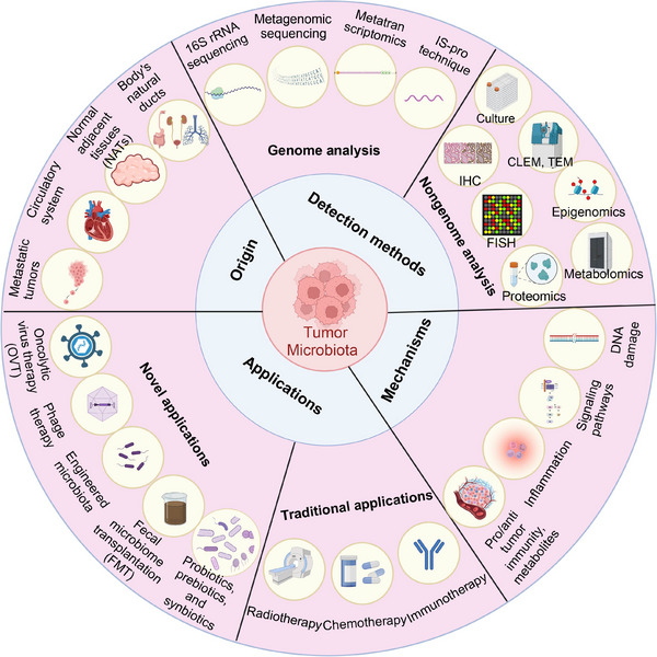
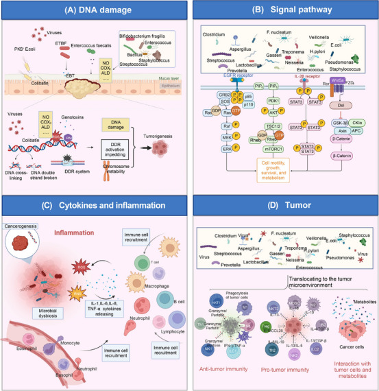
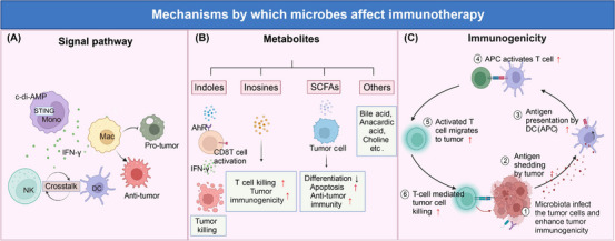
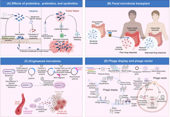
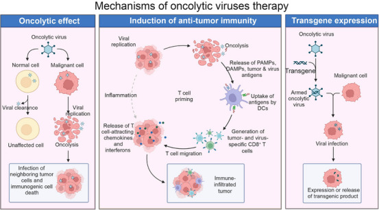
The intratumoral microbiome (TM) refers to the microorganisms in the tumor tissues, including bacteria, fungi, viruses, and so on, and is distinct from the gut microbiome and circulating microbiota. TM is strongly associated with tumorigenesis, progression, metastasis, and response to therapy. This paper highlights the current status of TM. Tract sources, adjacent normal tissue, circulatory system, and concomitant tumor co-metastasis are the main origin of TM. The advanced techniques in TM analysis are comprehensively summarized. Besides, TM is involved in tumor progression through several mechanisms, including DNA damage, activation of oncogenic signaling pathways (phosphoinositide 3-kinase [PI3K], signal transducer and activator of transcription [STAT], WNT/β-catenin, and extracellular regulated protein kinases [ERK]), influence of cytokines and induce inflammatory responses, and interaction with the tumor microenvironment (anti-tumor immunity, pro-tumor immunity, and microbial-derived metabolites). Moreover, promising directions of TM in tumor therapy include immunotherapy, chemotherapy, radiotherapy, the application of probiotics/prebiotics/synbiotics, fecal microbiome transplantation, engineered microbiota, phage therapy, and oncolytic virus therapy. The inherent challenges of clinical application are also summarized. This review provides a comprehensive landscape for analyzing TM, especially the TM-related mechanisms and TM-based treatment in cancer.

Keywords: analysis methods; immunotherapy; intratumoral microbiome; treatment application; tumor‐promotive and tumor‐suppressive mechanisms.


Conflict of interest statement

All authors declare that they have no competing interests.

Figures

FIGURE 1
FIGURE 1
An overview of the advances in TM. Illustrating the origin of TM (Tract sources, adjacent normal tissue, circulatory system, and concomitant tumor co‐metastasis), the TM analysis methods (16SrRNA sequencing, shotgun metagenomic sequencing, metatranscriptomics, IS‐pro technique, immunohistochemistry, fluorescence in situ hybridization, proteomics, metabolomics, correlative light and electron microscopy and transmission electron microscopy, TM Culture, single cell analysis and spatial transcriptome, organoids and 3D technology, gene chip technology, nanotechnology, computational tool, molecular detection method based on viral nucleic acid, immunological method, and nucleic acid hybridization), the potential mechanism of how TM is involved in tumors (DNA damage, activation of oncogenic signaling pathways, influence cytokines and induce inflammatory responses, and the interaction with tumor microenvironment, and the promising directions of TM‐based treatment (immunotherapy, chemotherapy, radiotherapy, the application of probiotics/prebiotics/synbiotics, fecal microbiome transplantation, engineered microbiota, phage therapy, and oncolytic virus therapy). Abbreviations: CLEM, correlative light and electron microscopy; FISH, fluorescence in situ hybridization; FMT, fecal microbiome transplantation; IHC, immunohistochemistry; OVT, Oncolytic virus therapy; TEM, transmission electron microscopy; TM, tumor microbiome (intratumoral microbiome). Biorender supported the materials in Figure 1.
FIGURE 2
FIGURE 2
Involvement of microbiota in cancer. Microbiota is involved in cancer and mainly affects carcinogenesis or cancer prevention through the four aspects. (A) Tumorigenesis of microbial damage to DNA. Microbiota dysbiosis is often related to tumor initiation. Pathogenic microorganisms produce more compounds (for example, NO, COX2, acetaldehyde) capable of causing DNA damage, chromosomal instability, impairment of the DNA damage response (DDR) system, etc., thus causing tumorigenesis. For instance, pks E. coli can invade the mucus layer and encode an unstable DNA alkylating agent, colibatin, causing DNA interstrand cross‐linking and double‐strand breaks. Other microbiota, including ETBF, Enterococcus faecallis, etc., secret genotoxins and lead to reactive oxygen species (ROS) and reactive nitrogen intermediates (RNI) production, eventually promoting DNA damage. (B) Activation of oncogenic signaling pathways. Microbes activate the PI3K signaling pathway, STAT signaling pathway, Wnt/β‐catenin signaling pathway, etc., influencing essential activities such as cell motility, growth, survival, and metabolism, resulting in carcinogenesis. (C) Inflammatory carcinogenesis caused by microbial dysbiosis. Microbial dysbiosis results in ROS and reactive nit ogen species (RNS) production, immune cell recruitment, and inflammatory microenvironment formation. Chronic inflammation promotes tumorigenesis and progression. (D) Interaction with the tumor microenvironment. Microorganisms translocate to the tumor microenvironment (TME), exerting pro‐tumor and anti‐tumor effects through enhancement of anti‐tumor immunity, suppression of anti‐tumor immunity, and interaction with tumor cells and metabolite production. Abbreviations: ALD, acetaldehyde; APC, antigen presenting cell; COX2, cyclooxygenase 2; DDR, DNA damage response; 3D, three‐dimensional; ETBF, enterotoxigenic Bifidobacterium fragilis; EBT, enterotoxigenic bacterial toxin; MDSC, marrow‐derived suppressor cell; PI3K, phosphoinositide 3 ‐ kinas; ROS, reactive oxygen species; RNI, reactive nitrogen intermediates; RNS, reactive nit rogen species; STAT, signal transducer and activator of transcription; TME, tumor microenvironment. Biorender supported the materials in Figure 2.
FIGURE 3
FIGURE 3
Mechanisms by which microbes affect immunotherapy. Microorganisms affect immunotherapy mainly in three ways: modulating signal pathways, producing metabolites, and enhancing immunogenicity. (A) modulating signal pathways: Microbiota‐derived agonists c‐di‐AMP modulate STING of monocyte, induce IFN‐γ secretion, bolster DC/NK cell crosstalk, and promote anti‐tumor macrophage activation. (B) metabolites producing: Many microbial metabolites, including indole, inosine, SCFAs, etc., have anti‐tumor effects. Indole activates CD8+ T cells via binding AhR and increases IFN‐γ releasing, thus enhancing tumor killing. Inosine promotes T‐cell killing and enhances tumor immunogenicity. Moreover, SCFAs inhibit tumor cell differentiation, induce apoptosis, and promote anti‐tumor response. (C) enhancing immunogenicity: Commensal microbiota increases tumor antigen shedding and antigen presentation, activating more T cells and migrating to the tumor, causing more tumor cell killing. Abbreviations: APC, antigen presenting cell; DC, dendritic cells; Mac, macrophage; NK, natural killer cell; SCFAs, short‐chain fatty acids; STING, stimulator of interferon genes. Biorender supported the materials in Figure 3.
FIGURE 4
FIGURE 4
Mechanisms of probiotics, fecal microbiome transplantation, engineered microbiota, and phage therapy. (A) Effect of probiotics. Prebiotics could promote the growth of probiotics. Probiotics colonize in the intestine and reshape the microflora. These beneficial microbiotas could regulate intestinal conditions (increasing anticancer substances and decreasing carcinogens). In addition, they also enhance the intestinal barrier function. Some probiotics enable translocation to tumor tissue and regulate signal pathways, inflammation, and metabolic conditions. (B) Fecal microbiome transplantation. Fecal microbiota from drug‐sensitive patients can overcome drug resistance and improve the anti‐tumor effects in patients with poor drug response through microbiota remodeling. (C) Engineered microbiota. The microbiota is engineered and equipped with the target gene and the nanoparticle coating layer. The engineered microbiota easily spreads into the bloodstream and the tumor tissue and then secretes target molecules that help kill tumors. (D) Phage display and phage vector. Phage display requires five steps: polypeptide‐displayed phage libraries generation and amplification, target molecule binding, removing unbound and nonspecific phages, elution of target‐bound phages and infection of host bacteria, and replication. These infective bacteria help in the early diagnosis, treatment, and prevention of cancer vaccines. Phages such as adeno‐associated viruses combined with therapeutic genes could enter the tumor cell and translocate nuclei into DNA. These genes enable the expression of toxins that cause apoptosis in tumor cells. Abbreviations: SCFAs, short‐chain fatty acids; FMT, fecal microbiome transplantation. Biorender supported the materials in Figure 4.
FIGURE 5
FIGURE 5
Mechanisms of oncolytic virus therapy. Oncolytic viruses (OVs) produce effects in two main ways. One is an oncolytic effect whereby OVs infect and replicate in selective tumor cells, eventually leading to tumor lysis. The other is that OVs aim at selective tumor cells and result in oncolysis, releasing viral and tumor‐associated antigens, danger‐associated molecular patterns (DAMPs), pathogen‐associated molecular patterns (PAMPs), T cell attracting cytokines and interferons, thereby recruiting and activating DCs, NK cells, and T cells, thus enhancing the anti‐tumor immunity. OVs also have a property that can express and release transgenic products as vectors in malignant cells. Abbreviations: DAMPs, danger‐associated molecular patterns; DC, dendritic cells; NK, natural killer cell; OVs, oncolytic viruses; PAMPs, pathogen‐associated molecular patterns. Biorender supported the materials in Figure 5.

References

    1. Nejman D, Livyatan I, Fuks G, Gavert N, Zwang Y, Geller LT, et al. The human tumor microbiome is composed of tumor type‐specific intracellular bacteria. Science. 2020;368(6494):973–980. - PMC - PubMed
    1. Lemmon MJ, van Zijl P, Fox ME, Mauchline ML, Giaccia AJ, Minton NP, et al. Anaerobic bacteria as a gene delivery system that is controlled by the tumor microenvironment. Gene Ther. 1997;4(8):791–796. - PubMed
    1. Yazawa K, Fujimori M, Amano J, Kano Y, Taniguchi S. Bifidobacterium longum as a delivery system for cancer gene therapy: selective localization and growth in hypoxic tumors. Cancer Gene Ther. 2000;7(2):269–274. - PubMed
    1. Alexeev EE, Lanis JM, Kao DJ, Campbell EL, Kelly CJ, Battista KD, et al. Microbiota‐Derived Indole Metabolites Promote Human and Murine Intestinal Homeostasis through Regulation of Interleukin‐10 Receptor. Am J Pathol. 2018;188(5):1183–1194. - PMC - PubMed
    1. Grivennikov S, Karin E, Terzic J, Mucida D, Yu GY, Vallabhapurapu S, et al. IL‐6 and Stat3 are required for survival of intestinal epithelial cells and development of colitis‐associated cancer. Cancer Cell. 2009;15(2):103–113. - PMC - PubMed