. 2024 Sep 11;15(9):e0038424.
doi: 10.1128/mbio.00384-24. Epub 2024 Aug 1.

Cytosolic serpins act in a cytoprotective feedback loop that limits ESX-1-dependent death of Mycobacterium marinum-infected macrophages

Affiliations

Cytosolic serpins act in a cytoprotective feedback loop that limits ESX-1-dependent death of Mycobacterium marinum-infected macrophages

Esther Nobs et al. mBio. .

Abstract

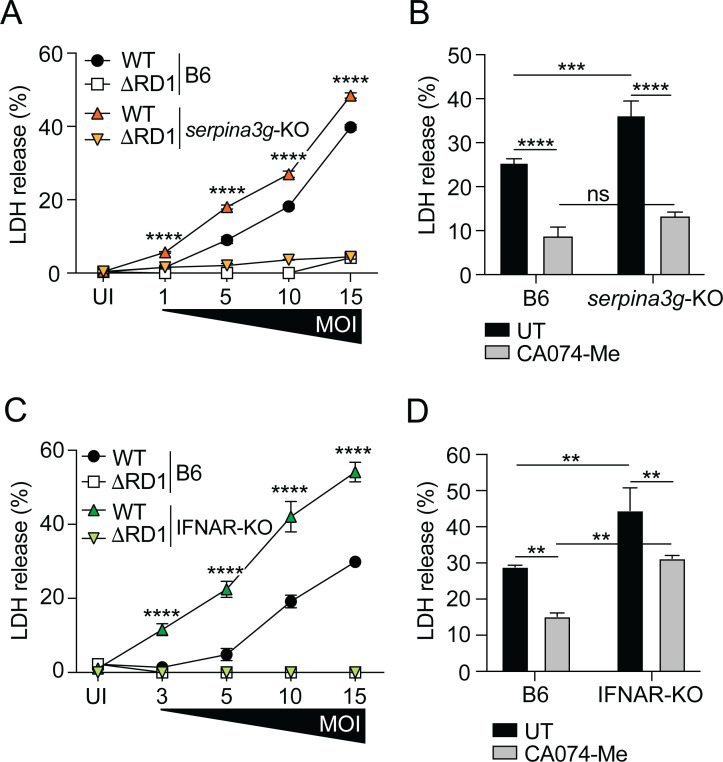
Serine protease inhibitors (serpins) constitute the largest family of protease inhibitors expressed in humans, but their role in infection remains largely unexplored. In infected macrophages, the mycobacterial ESX-1 type VII secretion system permeabilizes internal host membranes and causes leakage into the cytosol of host DNA, which induces type I interferon (IFN) production via the cyclic GMP-AMP synthase (cGAS) and stimulator of IFN genes (STING) surveillance pathway, and promotes infection in vivo. Using the Mycobacterium marinum infection model, we show that ESX-1-mediated type I IFN signaling in macrophages selectively induces the expression of serpina3f and serpina3g, two cytosolic serpins of the clade A3. The membranolytic activity of ESX-1 also caused leakage of cathepsin B into the cytosol where it promoted cell death, suggesting that the induction of type I IFN comes at the cost of lysosomal rupture and toxicity. However, the production of cytosolic serpins suppressed the protease activity of cathepsin B in this compartment and thus limited cell death, a function that was associated with increased bacterial growth in infected mice. These results suggest that cytosolic serpins act in a type I IFN-dependent cytoprotective feedback loop to counteract the inevitable toxic effect of ESX-1-mediated host membrane rupture.

Importance: The ESX-1 type VII secretion system is a key virulence determinant of pathogenic mycobacteria. The ability to permeabilize host cell membranes is critical for several ESX-1-dependent virulence traits, including phagosomal escape and induction of the type I interferon (IFN) response. We find that it comes at the cost of lysosomal leakage and subsequent host cell death. However, our results suggest that ESX-1-mediated type I IFN signaling selectively upregulates serpina3f and serpina3g and that these cytosolic serpins limit cell death caused by cathepsin B that has leaked into the cytosol, a function that is associated with increased bacterial growth in vivo. The ability to rupture host membranes is widespread among bacterial pathogens, and it will be of interest to evaluate the role of cytosolic serpins and this type I IFN-dependent cytoprotective feedback loop in the context of human infection.

Keywords: Spi2A; bacterial pathogenesis; cathepsin B; cytosolic surveillance pathways; host cell death; host-pathogen interactions; lysosome; membrane permeabilization; serpins; type I interferon; type VII secretion system.


Conflict of interest statement

The authors declare no conflict of interest.

Figures

Fig 1
Fig 1
ESX-1 induces type I IFN-dependent expression of cytosolic A3 serpins. (A) Schematic representation of the genetic region encoding A3 serpins in mice. Pseudogenes, not encoding a functional protein, are marked with an asterisk (30, 31), and serpina3f/g are highlighted in orange. serpina3f–i do not encode a signal peptide (30). (B, D, and E) Wild-type C57BL/6 (B6) and IFNAR-KO macrophages were infected with WT, ΔRD1, and ΔRD1::RD1 M. marinum at a multiplicity of infection (MOI) of 5, or left uninfected (UI), as indicated. (B) Kinetic reverse-transcription quantitative PCR (RT-qPCR) analysis gene expression, using primers against regions conserved among clade A3 serpin encoding genes. (C) B6 and IFNAR-KO mice were infected with WT M. marinum. At 14 days post-infection, the infected tail tissue was analyzed by RT-qPCR for the expression of A3 serpins as described for panel B above. Shown is the mean of four infected mice (and one uninfected mouse) per genotype, as indicated. Two-tailed unpaired t-test, **P < 0.01. (D) Expression of the individual A3 serpin encoding genes at 7 hpi. (E) RT-qPCR analysis of serpina3f and serpina3g expression at 7 hpi. Results (mean ± standard deviation [SD]; n = 3) are representative of three independent experiments. One-way analysis of variance (ANOVA), ****P < 0.0001. (B and D) Results (mean ± SD; n = 3) are representative of three independent experiments. Two-way ANOVA, ****P < 0.0001.
Fig 2
Fig 2
Cytosolic serpins inhibit the activity of extralysosomal cathepsin B. Macrophages were infected with WT and ΔRD1 M. marinum, or uninfected (UI), as indicated. (A) At 7 hpi, cathepsin B activity was measured in whole cell lysate, cytosolic fraction, and supernatant, as indicated. (B) Analysis of cathepsin B activity in the supernatant of WT-infected macrophages treated with inhibitors of cathepsin B (CA074-Me; 25 µM), cysteine proteases (E64; 10 µM), aspartyl proteases (pepstatin A; 10 µM). or cathepsin L (RKLLW-NH2; 10 µM). Untreated (UT) supernatants were analyzed as controls. (C) Kinetic RT-qPCR analysis of cathepsin B expression in macrophages infected (MOI = 5) as indicated. (D) Enzyme-linked immunosorbent assay-based analysis of cathepsin B protein concentration in the cytosolic fraction of WT-infected B6 and IFNAR-KO macrophages. Results (mean ± SD; n = 4) are representative of two independent experiments. Two-way ANOVA. (E) RT-qPCR analysis of the expression of the individual A3 serpin encoding genes in WT-infected B6 and serpina3g-KO macrophages (MOI = 5) at 7 hpi, as indicated. Uninfected (UI) cells were analyzed as controls. (F) Kinetic RT-qPCR analysis of cathepsin B expression in macrophages infected (MOI = 5) as indicated. (G) Analysis of cathepsin B activity in the supernatant of WT-infected B6 and serpina3g-KO macrophages, as indicated. (A–C and E–G) Results (mean ± SD; n = 3) are representative of three independent experiments. Two-way ANOVA, ****P < 0.0001.
Fig 3
Fig 3
Cytosolic serpins do not regulate inflammasome activation in M. marinum-infected macrophages. (A and B) B6 and serpina3g-KO macrophagesmacrophages were infected with WT M. marinum (MOI = 10) and treated with the cathepsin B inhibitor CA074-Me (25 µM), as indicated. The concentration of IL-1β (A) and IL-6 (B) secreted into the supernatant was determined by enzyme-linked immunosorbent assay (ELISA) at 24 hpi. (C and D) B6 and serpina3g-KO macrophages were infected with WT and ΔRD1 M. marinum at titrated MOI, as indicated. The concentration of IL-1β (C) and IL-6 (D) secreted into the supernatant was determined by ELISA at 24 hpi. (A–D) Results (mean ± SD; n = 3) are representative of three independent experiments (two-way ANOVA, ****P < 0.0001).
Fig 4
Fig 4
Cytosolic serpins inhibit ESX-1-dependent host cell death. B6, IFNAR-KO, and serpina3g-KO macrophages were infected with WT and ΔRD1 M. marinum, or uninfected (UI), as indicated. (A) LDH in the supernatant of B6 and serpina3g-KO macrophages infected at increasing MOI was measured at 7 hpi. (B) WT-infected macrophages (MOI = 10) were treated with the cathepsin B inhibitor CA074-Me (25 µM), and LDH release into the supernatant was measured at 7 hpi. (C) LDH in the supernatant of B6 and IFNAR-KO macrophages infected at increasing MOI was measured at 7 hpi. (D) WT-infected macrophages (MOI = 10) were treated with CA074-Me (25 µM), and LDH release into the supernatant was measured at 7 hpi. (A–D) Results (mean ± SD; n = 3) are representative of three independent experiments. Two-way ANOVA, ****P < 0.0001.
Fig 5
Fig 5
Cytosolic serpins promote M. marinum growth in vivo. B6 and serpina3g-KO mice were infected with WT M. marinum (1.4 × 10⁷ CFUs) via tail vein injection. (A) Kinetic analysis of the accumulated length (mm) of visible tail lesions. Each symbol indicates an individual mouse (n = 11 per group), and the bars show the mean for each group. Two-way ANOVA. (B and C) Bacterial burden in tail tissue and tail-draining (sciatic and inguinal, pooled) lymph nodes (LN) at 14 and 28 days post-infection (DPI), as indicated. Results (n = 9–15 mice per group) from two independent experiments. Bars indicate the mean for each group. Two-tailed unpaired t-test; *P < 0.05, **P < 0.01.
Fig 6
Fig 6
Working model. ESX-1-dependent permeabilization of host membranes causes leakage of host DNA (illustrated with a mitochondrion) into the cytosol and induces type I IFN via the cGAS-STING pathway. Type I IFN signaling leads to expression of the cytosolic serpins serpina3f and serpina3g, which inhibits cathepsin B as it reaches the cytosol due to ESX-1-dependent lysosomal rupture. Inhibition of cytosolic cathepsin B by serpins reduces the level of cell death in vitro and is associated with a higher bacterial burden in vivo.

References

    1. Stinear TP, Seemann T, Harrison PF, Jenkin GA, Davies JK, Johnson PDR, Abdellah Z, Arrowsmith C, Chillingworth T, Churcher C, et al. . 2008. Insights from the complete genome sequence of Mycobacterium marinum on the evolution of Mycobacterium tuberculosis. Genome Res 18:729–741. doi:10.1101/gr.075069.107 - DOI - PMC - PubMed
    1. Wolf AJ, Linas B, Trevejo-Nuñez GJ, Kincaid E, Tamura T, Takatsu K, Ernst JD. 2007. Mycobacterium tuberculosis infects dendritic cells with high frequency and impairs their function in vivo. J Immunol 179:2509–2519. doi:10.4049/jimmunol.179.4.2509 - DOI - PubMed
    1. Lienard J, Munke K, Wulff L, Da Silva C, Vandamme J, Laschanzky K, Joeris T, Agace W, Carlsson F. 2023. Intragranuloma accumulation and inflammatory differentiation of neutrophils underlie mycobacterial ESX-1-dependent immunopathology. mBio 14:e0276422. doi:10.1128/mbio.02764-22 - DOI - PMC - PubMed
    1. Cohen SB, Gern BH, Delahaye JL, Adams KN, Plumlee CR, Winkler JK, Sherman DR, Gerner MY, Urdahl KB. 2018. Alveolar macrophages provide an early Mycobacterium tuberculosis niche and initiate dissemination. Cell Host Microbe 24:439–446. doi:10.1016/j.chom.2018.08.001 - DOI - PMC - PubMed
    1. Hsu T, Hingley-Wilson SM, Chen B, Chen M, Dai AZ, Morin PM, Marks CB, Padiyar J, Goulding C, Gingery M, Eisenberg D, Russell RG, Derrick SC, Collins FM, Morris SL, King CH, Jacobs WR. 2003. The primary mechanism of attenuation of bacillus Calmette-Guérin is a loss of secreted lytic function required for invasion of lung interstitial tissue. Proc Natl Acad Sci U S A 100:12420–12425. doi:10.1073/pnas.1635213100 - DOI - PMC - PubMed

MeSH terms