. 2024 Aug 28;9(8):e0008924.
doi: 10.1128/msphere.00089-24. Epub 2024 Aug 1.

Synergistic effects of novel penicillin-binding protein 1A amino acid substitutions contribute to high-level amoxicillin resistance of Helicobacter pylori

Affiliations

Synergistic effects of novel penicillin-binding protein 1A amino acid substitutions contribute to high-level amoxicillin resistance of Helicobacter pylori

Alain Cimuanga-Mukanya et al. mSphere. .

Abstract

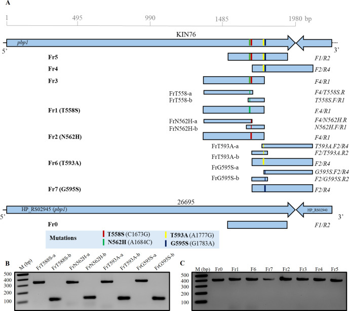
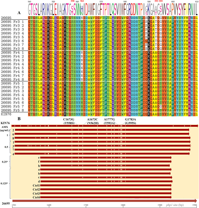
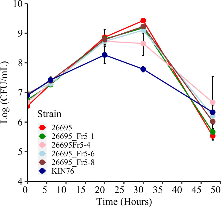
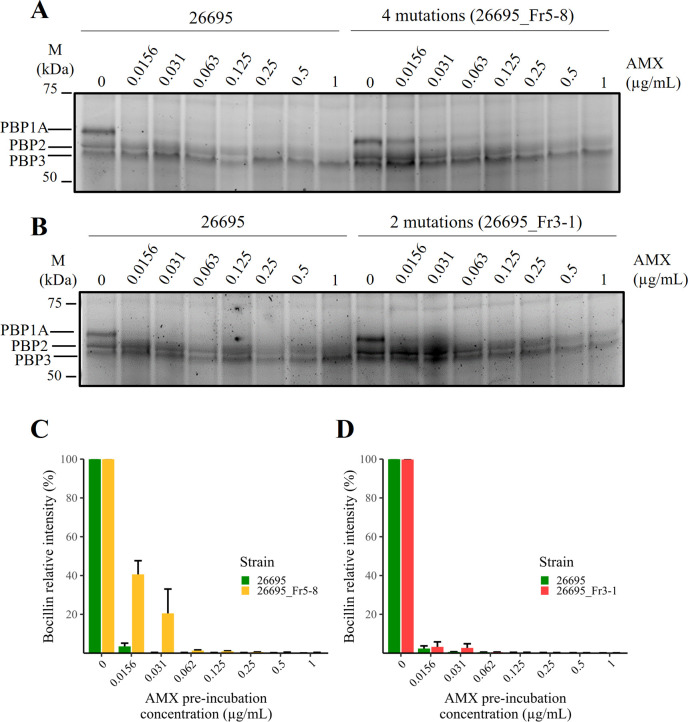
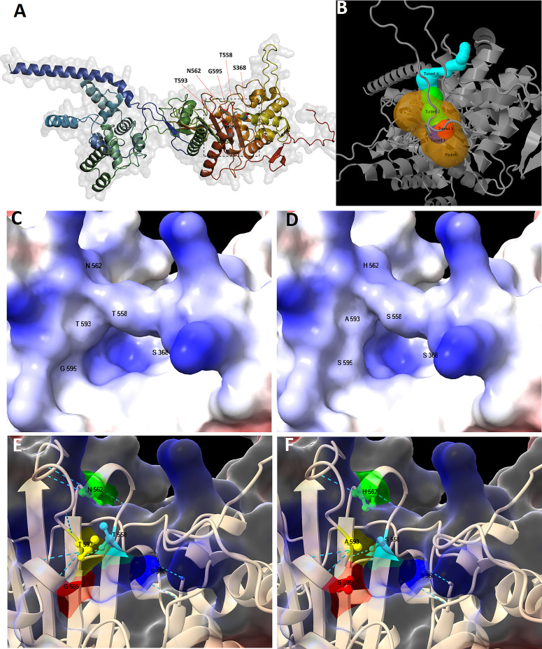
The growing resistance to amoxicillin (AMX)-one of the main antibiotics used in Helicobacter pylori eradication therapy-is an increasing health concern. Several mutations of penicillin-binding protein 1A (PBP1A) are suspected of causing AMX resistance; however, only a limited set of these mutations have been experimentally explored. This study aimed to investigate four PBP1A mutations (i.e., T558S, N562H, T593A, and G595S) carried by strain KIN76, a high-level AMX-resistant clinical H. pylori isolate with an AMX minimal inhibition concentration (MIC) of 2 µg/mL. We transformed a recipient strain 26695 with the DNA containing one to four mutation allele combinations of the pbp1 gene from strain KIN76. Transformants were subjected to genomic exploration and antimicrobial susceptibility testing. The resistance was transformable, and the presence of two to four PBP1A mutations (T558S and N562H, or T593A and G595S), rather than separate single mutations, was necessary to synergistically increase the AMX MIC up to 16-fold compared with the wild-type (WT) strain 26695. An AMX binding assay of PBP1A was performed using these strains, and binding was visualized by chasing Bocillin, a fluorescent penicillin analog. This revealed that all four-mutation allele-transformed strains exhibited decreased affinity to AMX on PBP1A than the WT. Protein structure modeling indicated that functional modifications occur as a result of these amino acid substitutions. This study highlights a new synergistic AMX resistance mechanism and establishes new markers of AMX resistance in H. pylori.IMPORTANCEThe development of resistance to antibiotics, including amoxicillin, is hampering the eradication of Helicobacter pylori infection. The identification of mechanisms driving this resistance is crucial for the development of new therapeutic strategies. We have demonstrated in vitro the synergistic role of novel mutations in the pbp1 gene of H. pylori that is suspected to drive amoxicillin resistance. Also deepening our understanding of amoxicillin resistance mechanisms, this study establishes new molecular markers of amoxicillin resistance that may be useful in molecular-based antibiotic susceptibility testing approaches for clinical practice or epidemiologic investigations.

Keywords: Helicobacter pylori; amoxicillin resistance; mutations; penicillin-binding protein 1A.


Conflict of interest statement

The authors declare no conflict of interest.

Figures

Fig 1
Fig 1
Amplicon construction. The pbp1 DNA fragments carrying different rearrangements of mutations were amplified by PCR from genomic DNA of clinical isolate KIN76 (A). Amplicons Fr1, Fr2, Fr6, and Fr7 carrying a single mutation were additionally constructed by fusion PCR of the single-stranded DNA fragments FrT558S-a/FrT558S-b, Fr2a/Frb2, FrT593A-a/FrT593A-b, and G595S-a/G595S-b, respectively. A DNA fragment (Fr0) without any mutation was also prepared from 26695 genomic DNA and used as a negative control. The PCR fragments before fusion PCR (B). All the final KIN76 amplicons prepared before transformation into strain 26695 (C). All the PCR fragments were checked in 1.8% agarose S gel electrophoresis.
Fig 2
Fig 2
Alignment of PBP1A amino acid and pbp1 nucleotide sequences. (A) Alignment of PBP1A amino acid sequences. After Sanger sequencing and initial processing of the raw data, the pbp1 gene fragments from transformants were aligned with sequences from the WT 26695 strain and the KIN76 clinical strain, then translated into protein sequences. T558 and N562 of strain 26695 were substituted by S and H in all transformant strains obtained by transformation with the Fr3 DNA fragment, except one strain (26695_Fr3-6) that contained not-AMX-R-related G589, same as strain KIN76. T593 and G595 of strain 26695 were substituted by A and S in transformant strains obtained by transformation with the Fr4 DNA fragment in all strains. Similarly, in transformants resulting from transformation with the Fr5 DNA fragment, substitutions occurred on all four loci in every strain sequenced except for one strain (26695_Fr5-7) that showed a conserved G595. (B) Nucleotide sequences of Fr5 and control strains. All the Fr5 strains (Fr5-1 to Fr5-8) were transformed using the Fr5 amplicon, and their MICs, determined later, ranged from 0.25 to 1 µg/mL. The asterisk indicates that each colony of the strain was selected on a plate containing AMX at the indicated concentration. Sequences Fr5-05 (e, f, and h) derive from strains selected on plates containing 0.5 µg/mL AMX, whereas sequences Fr5-0125 (a–d) are from Fr5-transformant strains picked from non-selective plates with 0.125 µg/mL AMX. Control sequences (Ctrl1 to Ctrl4) are from control colonies picked from non-selective plates. All sequences were aligned to the pbp1 gene of WT strains 26695 and KIN76, spanning nucleotides 1,501 to 1,980. Vertical lines within the sequences denote nucleotide variation sites in comparison to the 26695 sequence. Mutation sites are indicated at the top of the figure.
Fig 3
Fig 3
Growth rate of WT and Fr5 mutant strains. This figure depicts the growth rates of the WT strain KIN76 and the AMX-R mutant strains 26695_Fr5-1, 26695_Fr5-4, 26695_Fr5-6, and 26695_Fr5-8 compared to the susceptible strain 26695. Colony-forming units (CFUs) were counted at 0, 6, 20, 30, 48, and 72 hours post-inoculation. The results are presented as the means and standard errors of four independent experiments. The mutant strains did not exhibit a significant difference from the WT 26695, whereas the growth rate of strain KIN76 showed a significant difference compared to 26695 by the Wilcoxon signed-rank test (P = 0.0264).
Fig 4
Fig 4
AMX-Bocillin competitive binding assay. The relative signal intensity of Bocillin was set to maximal when no AMX was added to the binding assay reaction (AMX preincubation concentration = 0). However, preincubation with 0.0156–0.031 µg/mL of AMX showed a higher Bocillin signal in transformant 26695_Fr5-8 (A, C) than transformant 26695_Fr3-8 (B, D), indicating a decrease in PBP1A affinity for AMX in transformants carrying mutations within PBP1A, with a greater effect seen with PBP1A harboring four mutations than PBP1A harboring only two mutations. The total protein intensity for each lane (not shown) was used for Bocillin quantification. Three independent assays were performed (n = 3).
Fig 5
Fig 5
H. pylori PBP1A tertiary structure models. Whole structure of PBP1A of strain 26695, modeled through Alfafold2Colab and visualized using PyMol (A); the square roughly indicates the pocket, and the red arrows indicate residues at positions 368, 558, 562, 593, and 595. The pocket with the highest relevance score, centered around catalytic residue S368 (hidden in the figure), and the corresponding tunnels as predicted by Caver (B). Closed-up structures of PBP1A near the catalytic residue were visualized without mutation (C, E) and with four mutations (D, F) using CSF-Chimera. In surface models (C, D), regions of positive charge are shaded blue (basic), and those of negative charge are shaded red (acidic). In ribbon models (E, F), residues in positions 558 (light green), 562 (orange yellow), 593 (orange), and 595 (red) are shown to line to the access route to catalytic residue S368 (blue). The narrow green line traces the approximate contour of the residues, and dotted lines correspond to hydrogen bonds.

Similar articles

Cited by

References

    1. Dang BN, Graham DY. 2017. Helicobacter pylori infection and antibiotic resistance: a WHO high priority? Nat Rev Gastroenterol Hepatol 14:383–384. doi:10.1038/nrgastro.2017.57 - DOI - PMC - PubMed
    1. Salama NR, Hartung ML, Müller A. 2013. Life in the human stomach: persistence strategies of the bacterial pathogen Helicobacter pylori. Nat Rev Microbiol 11:385–399. doi:10.1038/nrmicro3016 - DOI - PMC - PubMed
    1. Sanders MK, Peura DA. 2002. Helicobacter pylori-associated diseases. Curr Gastroenterol Rep 4:448–454. doi:10.1007/s11894-002-0019-x - DOI - PubMed
    1. Munita JM, Arias CA. 2016. Mechanisms of antibiotic resistance. Microbiol Spectr 4. doi:10.1128/microbiolspec.VMBF-0016-2015 - DOI - PMC - PubMed
    1. Thung I, Aramin H, Vavinskaya V, Gupta S, Park JY, Crowe SE, Valasek MA. 2016. Review article: the global emergence of Helicobacter pylori antibiotic resistance. Aliment Pharmacol Ther 43:514–533. doi:10.1111/apt.13497 - DOI - PMC - PubMed

MeSH terms

LinkOut - more resources