. 2024 Oct;119(5):733-749.
doi: 10.1007/s00395-024-01071-z. Epub 2024 Aug 1.

SGLT1 contributes to glucose-mediated exacerbation of ischemia-reperfusion injury in ex vivo rat heart

Affiliations

SGLT1 contributes to glucose-mediated exacerbation of ischemia-reperfusion injury in ex vivo rat heart

Alhanoof Almalki et al. Basic Res Cardiol. 2024 Oct.

Abstract

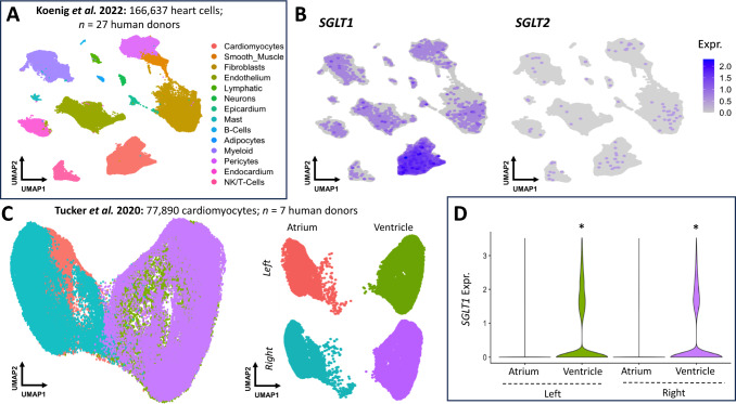
Hyperglycaemia is common during acute coronary syndromes (ACS) irrespective of diabetic status and portends excess infarct size and mortality, but the mechanisms underlying this effect are poorly understood. We hypothesized that sodium/glucose linked transporter-1 (SGLT1) might contribute to the effect of high-glucose during ACS and examined this using an ex-vivo rodent heart model of ischaemia-reperfusion injury. Langendorff-perfused rat hearts were subjected to 35 min ischemia and 2 h reperfusion, with variable glucose and reciprocal mannitol given during reperfusion in the presence of pharmacological inhibitors of SGLT1. Myocardial SGLT1 expression was determined in rat by rtPCR, RNAscope and immunohistochemistry, as well as in human by single-cell transcriptomic analysis. High glucose in non-diabetic rat heart exacerbated reperfusion injury, significantly increasing infarct size from 45 ± 3 to 65 ± 4% at 11-22 mmol/L glucose, respectively (p < 0.01), an association absent in diabetic heart (32 ± 1-37 ± 5%, p = NS). Rat heart expressed SGLT1 RNA and protein in vascular endothelium and cardiomyocytes, with similar expression found in human myocardium by single-nucleus RNA-sequencing. Rat SGLT1 expression was significantly reduced in diabetic versus non-diabetic heart (0.608 ± 0.08 compared with 1.116 ± 0.13 probe/nuclei, p < 0.01). Pharmacological inhibitors phlorizin, canagliflozin or mizagliflozoin in non-diabetic heart revealed that blockade of SGLT1 but not SGLT2, abrogated glucose-mediated excess reperfusion injury. Elevated glucose is injurious to the rat heart during reperfusion, exacerbating myocardial infarction in non-diabetic heart, whereas the diabetic heart is resistant to raised glucose, a finding which may be explained by lower myocardial SGLT1 expression. SGLT1 is expressed in vascular endothelium and cardiomyocytes and inhibiting SGLT1 abrogates excess glucose-mediated infarction. These data highlight SGLT1 as a potential clinical translational target to improve morbidity/mortality outcomes in hyperglycemic ACS patients.

Keywords: Diabetes; Hyperglycemia; Myocardial infarction; Reperfusion injury; SGLT1; SGLT2.


Conflict of interest statement

There are no conflicts of interest to declare.

Figures

Fig. 1
Fig. 1
Glucose-infarct size in non-diabetic and diabetic mouse and rat heart models. A Protocol for ex-vivo experiments. During reperfusion, mannitol is used to ensure that osmolality across different glucose concentration groups remains the same, thus with higher glucose concentration, the mannitol concentration is reciprocally lower. B C57BL/6 J non-diabetic mouse hearts (n = 4–5/group), subjected to global ischaemia, revealed a dose-range effect with minimum and maximum at 11 mmol/L and 22 mmol/L glucose, respectively. A linear relationship between glucose and infarct size was observed with incremental glucose, with an r2 = 0.35, p = 0.047. C SD non-diabetic rat hearts (n = 6/group), subjected to regional ischaemia, had minimum and maximum infarction at 11–22 mmol/L glucose, respectively, with a linear relationship between these values (r2 = 0.56, p = 0.0009). D GK diabetic rat heart (n = 6/group), subjected to regional ischaemia, had a negligible relationship between glucose and final infarct size when compared to non-diabetic hearts, with minimum infarction occurring at 16.5 mmol/L and attenuated peak infarct sizes
Fig. 2
Fig. 2
SGLT1 and SGLT2 expression in healthy rat and human myocardium. A Sglt1 and Sglt2 rtPCR in rat myocardium (left ventricle, LV), kidney (K, positive control) and skeletal muscle (SkM, negative control). In heart Sglt1 (using primers to detect exon 13–15), but not Sglt2 mRNA is detected (n = 5–9, representative blots shown). Gapdh: housekeeping gene, Glyceraldehyde-3-phosphate dehydrogenase. B Agarose gel electrophoresis depicting PCR amplification products for the rat Sglt1 gene using primers to detect exons 1–3, 3–5 and 8–9. DNA fragments of the expected size for Sglt1 are visible. Lane labels: G-gut, K-kidney, LV-left ventricle, each correspond to individual rat samples. Hprt: housekeeping gene, Hypoxanthine Guanine Phosphoribosyl Transferase
Fig. 3
Fig. 3
Human cellular expression of SGLT1. A Uniform manifold approximation and projection (UMAP) of snRNA-seq data of human heart cells from Koenig et al. 14 distinct cell types were identified, including cardiomyocytes. B SGLT1 expression (left UMAP) is enriched in cardiomyocytes. Conversely, SGLT2 expression (right UMAP) is scant. C UMAP of snRNA-seq data human cardiomyocytes (top UMAP) from Tucker et al. The bottom UMAP represents stratification of cardiomyocytes into the four chambers, with atrial and ventricular cardiomyocytes resolving separately. Analysis of the relative expression of SGLT1 in atrial versus ventricular cardiomyocytes (D)
Fig. 4
Fig. 4
SGLT1 and SGLT2 expression in the SD rat myocardium. A RNAscope revealed Sglt1 mRNA expression in both cardiomyocytes (which auto-fluoresce red), and vascular structures. Kidney is a positive control, with a strong SGLT1 signal (red) within the proximal nephron. v ventricular cardiomyocyte, ec endothelial cell, rbc red blood cell, tc tubular cell. Yellow arrows highlight SGLT detection signal: here predominantly in DAPI stained nuclei showing red/magenta signal. B There was no SGLT2 signal detectable within the heart, in contrast to kidney. Vascular markers, cell membrane marker, WGA (green) (C), and endothelial marker CD31 (green) (D) were used to show SGLT1 distribution. E Immunohistochemistry for SGLT1 protein expression (brown) reveals similar cellular staining pattern in heart and kidney to that seen with RNAscope. In the heart, staining can be found in the sarcolemma of the cardiomyocytes and the endothelium of arterioles and capillary structures (highlighted with red arrows)
Fig. 5
Fig. 5
Inhibition of SGLT2 and SGLT1 in 11 and 22 mmol/L glucose at reperfusion. A Chemical structures of the SGLT inhibitors used. B In non-diabetic SD rat heart, elevated glucose at reperfusion results in a significant increase in infarct over size the control condition which is completely abrogated by the administration of phlorizin (3 µmol/L). **p < 0.001 22 mmol/L versus 11 mmol/L reperfusion glucose. C There is a significant increase in infarct size with high (22 mmol/L) glucose versus 11 mmol/L, which is not ameliorated by 5 nmol/L canagliflozin. However, 1 µmol/L canagliflozin abrogates this excess injury. D Mizagliflozin, abrogates the excess injury associated with elevated glucose. Representative mid-ventricular myocardial slices shown. *p < 0.05 versus respective control
Fig. 6
Fig. 6
SGLT1 and SGLT2 expression in non-diabetic and diabetic heart. A In ZL rat heart, myocardial Sglt1 mRNA expression was markedly higher than that seen in ZDF rat heart (n = 6/group). B Sglt1 mRNA expression in kidney was not significantly altered in the ZDF diabetic model. C Diabetes did not result in a compensatory increase of Sglt2 mRNA expression. D Renal Sglt2 expression is significantly increased in diabetic kidney versus non-diabetic kidney. *p < 0.05

References

    1. Abbott GW, Tai KK, Neverisky DL, Hansler A, Hu Z, Roepke TK, Lerner DJ, Chen Q, Liu L, Zupan B, Toth M, Haynes R, Huang X, Demirbas D, Buccafusca R, Gross SS, Kanda VA, Berry GT (2014) KCNQ1, KCNE2, and Na+-coupled solute transporters form reciprocally regulating complexes that affect neuronal excitability. Sci Signal. 10.1126/scisignal.2005025 - PMC - PubMed
    1. Andreadou I, Bell RM, Botker HE, Zuurbier CJ (2020) SGLT2 inhibitors reduce infarct size in reperfused ischemic heart and improve cardiac function during ischemic episodes in preclinical models. Biochim Biophys Acta Mol Basis Dis 1866:165770. 10.1016/j.bbadis.2020.165770 - PubMed
    1. Banerjee SK, McGaffin KR, Pastor-Soler NM, Ahmad F (2009) SGLT1 is a novel cardiac glucose transporter that is perturbed in disease states. Cardiovasc Res 84:111–118. 10.1093/cvr/cvp190 - PMC - PubMed
    1. Barquera S, Pedroza-Tobias A, Medina C, Hernandez-Barrera L, Bibbins-Domingo K, Lozano R, Moran AE (2015) Global overview of the epidemiology of atherosclerotic cardiovascular disease. Arch Med Res 46:328–338. 10.1016/j.arcmed.2015.06.006 - PubMed
    1. Bell RM, Mocanu MM, Yellon DM (2011) Retrograde heart perfusion: the langendorff technique of isolated heart perfusion. J Mol Cell Cardiol 50:940–950. 10.1016/j.yjmcc.2011.02.018 - PubMed

Publication types

MeSH terms

LinkOut - more resources