

Utility of pan-bacterial and pan-fungal PCR in endophthalmitis: case report and review of the literature
- PMID: 39088113
- PMCID: PMC11294505
- DOI: 10.1186/s12348-024-00419-9
Utility of pan-bacterial and pan-fungal PCR in endophthalmitis: case report and review of the literature
Abstract
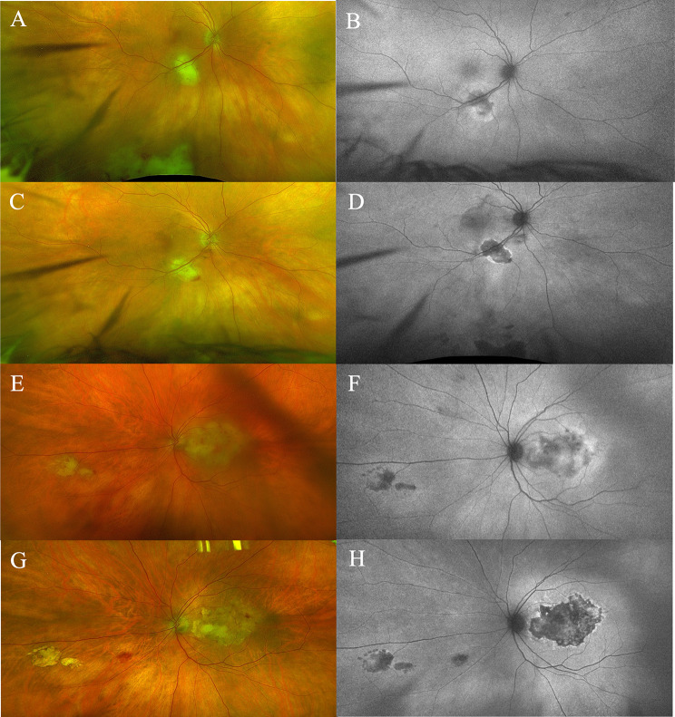
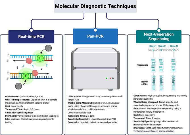
Background: Endophthalmitis is a clinical diagnosis but identification of the disease-causing agent or agents allows for a more tailored treatment. This is routinely done through intraocular fluid cultures and staining. However, culture-negative endophthalmitis is a relatively common occurrence, and a causative organism cannot be identified. Thus, further diagnostic testing, such as pan-bacterial and pan-fungal polymerase chain reactions (PCRs), may be required. BODY: There are now newer, other testing modalities, specifically pan-bacterial and pan-fungal PCRs, that may allow ophthalmologists to isolate a causative agent when quantitative PCRs and cultures remain negative. We present a case report in which pan-fungal PCR was the only test, amongst quantitative PCRs, cultures, and biopsies, that was able to identify a pathogen in endophthalmitis. Pan-PCR has unique advantages over quantitative PCR in that it does not have a propensity for false-positive results due to contamination. Conversely, pan-PCR has drawbacks, including its inability to detect viruses and parasites and its increased turnaround time and cost. Based on two large retrospective studies, pan-PCR was determined not to be recommended in routine cases of systemic infection as it does not typically add value to the diagnostic workup and does not change the treatment course in most cases. However, in cases like the one presented, pan-bacterial and pan-fungal PCRs may be considered if empiric treatment fails or if the infective organism cannot be isolated. If pan-PCR remains negative or endophthalmitis continues to persist, an even newer form of testing, next-generation sequencing, may aid in the diagnostic workup of culture-negative endophthalmitis.
Conclusion: Pan-bacterial and pan-fungal PCR testing is a relatively new diagnostic tool with unique advantages and drawbacks compared to traditional culturing and PCR methods. Similar to the tests' use in non-ophthalmic systemic infections, pan-bacterial and pan-fungal PCRs are unlikely to become the initial diagnosis test and completely replace culture methods. However, they can provide useful diagnostic information if an infectious agent is unable to be identified with traditional methods or if empiric treatment of endophthalmitis continues to fail.
Keywords: Chorioretinitis; Endophthalmitis; Intraocular infection; Next-generation sequencing; Quantitative PCR; pan-PCR.
© 2024. The Author(s).
Conflict of interest statement
The authors declare no competing interests.
Figures

References
Publication types
LinkOut - more resources
Full Text Sources
