. 2024 Sep 2;39(9):1240-1252.
doi: 10.1093/jbmr/zjae124.

Loss of the long form of Plod2 phenocopies contractures of Bruck syndrome-osteogenesis imperfecta

Affiliations

Loss of the long form of Plod2 phenocopies contractures of Bruck syndrome-osteogenesis imperfecta

Alexander Kot et al. J Bone Miner Res. .

Abstract

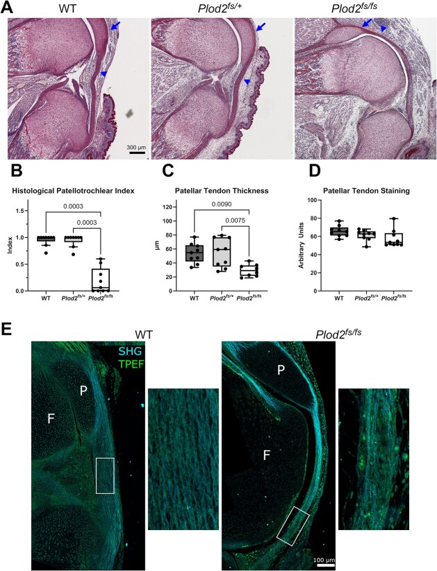
Bruck syndrome is an autosomal recessive form of osteogenesis imperfecta caused by biallelic variants in PLOD2 or FKBP10 and is characterized by joint contractures, bone fragility, short stature, and scoliosis. PLOD2 encodes LH2, which hydroxylates type I collagen telopeptide lysines, a critical step for collagen crosslinking. The Plod2 global knockout mouse model is limited by early embryonic lethality, and thus, the role of PLOD2 in skeletogenesis is not well understood. We generated a novel Plod2 mouse line modeling a variant identified in two unrelated individuals with Bruck syndrome: PLOD2 c.1559dupC, predicting a frameshift and loss of the long isoform LH2b. In the mouse, the duplication led to loss of LH2b mRNA as well as significantly reduced total LH2 protein. This model, Plod2fs/fs, survived up to E18.5 although in non-Mendelian genotype frequencies. The homozygous frameshift model recapitulated the joint contractures seen in Bruck syndrome and had indications of absent type I collagen telopeptide lysine hydroxylation in bone. Genetically labeling tendons with Scleraxis-GFP in Plod2fs/fs mice revealed the loss of extensor tendons in the forelimb by E18.5, and developmental studies showed extensor tendons developed through E14.5 but were absent starting at E16.5. Second harmonic generation showed abnormal tendon type I collagen fiber organization, suggesting structurally abnormal tendons. Characterization of the skeleton by μCT and Raman spectroscopy showed normal bone mineralization levels. This work highlights the importance of properly crosslinked type I collagen in tendon and bone, providing a promising new mouse model to further our understanding of Bruck syndrome.

Keywords: Bruck syndrome; LH2; PLOD2; bone; collagen crosslinking; hydroxylysine; mouse model; osteogenesis imperfecta; tendon.

Plain language summary

Bruck syndrome is a rare disease where individuals have brittle bone as well as contracted or stiff joints. Mutations in two genes are associated with Bruck syndrome and, in this work, we focus on PLOD2. Mice without Plod2 die at an early embryonic stage, before they have a chance to fully develop. In this work, we created a mouse with a PLOD2 mutation seen in people with Bruck syndrome. Some of these new Bruck syndrome model mice survived to a later gestational age, but all died at birth. The Bruck syndrome mice were small and had contracted joints. We found that they were missing tendons in their arms and had structurally abnormal tendons in their knees. Bone mineralization was normal, but there were indications that the modifications needed for normal type I collagen structure were absent. Overall, this is an advantageous new mouse model of Bruck syndrome that can be used to study this rare disease and highlights the importance of Plod2 in tendon.


Conflict of interest statement

The authors have no conflicts of interest to disclose.

Figures

Figure 1
Figure 1
The Plod2 frameshift allele specifically reduced LH2b levels and Plod2fs/fs mice recapitulate Bruck syndrome contractures. (A) Genetic strategy to model the duplication variant. Homologous recombination was used to insert the frameshift variant (red *) into the mouse genome. The frameshift allele contains LoxP sites, which were used for genotyping (arrows indicate primer locations). (B) Embryos were isolated from Plod2fs/+ x Plod2fs/+ crosses at multiple developmental ages. p-values shown from chi-squared tests when compared with expected mendelian ratios. Number of animals for each genotype analyzed noted in parentheses. (C) E18.5 Plod2fs/fs mice had contractures in all limbs (arrows) and altered cervical spine curvature (arrowhead). Heterozygous mice were indistinguishable from WT littermates. Lateral X-ray images at E18.5 demonstrated loss of the normal cervical spine curvature (arrow) at E18.5. Skeletal preparations revealed normal skeletal patterning. Plod2fs/fs mice were extremely delicate and disarticulated during the skeletal preparation protocol. Scale bars 0.5 cm. (D, E) Western blot analysis and quantification of relative LH2 levels in E18.5 calvaria lysates. n = 8/genotype. (F) qPCR analysis of the short form LH2a levels, n = 8 WT, 7 Plod2fs/+, and 8 Plod2fs/fs. (G) qPCR analysis of the long form LH2b levels, n = 8/genotype. Quantifications displayed with median and interquartile range.
Figure 2
Figure 2
Type I collagen C-telopeptide cross-linking lysine was not hydroxylated in E18.5 Plod2fs/fs mouse bone. (A, B) LC–MS profiles of in-gel trypsin digests of the α1(I) chain from WT and Plod2fs/fs E18.5 mouse bone. (C, D) MS/MS fragmentation spectrum of the parent ions from WT (1240.33+) and Plod2fs/fs (1240.43+) mouse collagen (black arrows). The sequence is shown with b and y ion breakages. P*, hydroxyproline; K*, hydroxylysine.
Figure 3
Figure 3
Loss of extensor tendons in Plod2fs/fs mice lead to forelimb contractures. (A) The Plod2fs line crossed with Scleraxis-GFP enabled the visualization of GFP+ tendon structures (dorsal view). At E18.5, the extensor carpi radialis longus/brevis, extensor digitorium communis, and extensor digiti quarti/quinti tendons were absent in the homozygous mouse forelimb (arrows). (B) Forelimbs were dissected from E18.5 homozygous mice and imaged, then tendons along the palmar side of the wrist were transected and the wrists were gently straightened with forceps. Contractures resolved after transection of flexor tendons. (C–E) Picrosirius red stained forelimb cross sections from E14.5, E16.5, and E18.5 mice. The 13 forelimb tendons were identified based on Watson et al. 2009. Multiple tendons formed (labeled and arrows) and were progressively absent (red boxes) in Plod2fs/fs mice. E14.5, n = 3 WT, 4 Plod2fs/+, and 3 Plod2fs/fs; E16.5 n = 4 WT, 5 Plod2fs/+, and 5 Plod2fs/fs; and E18.5 n = 9 WT, 6 Plod2fs/+, and 4 Plod2fs/fs. Radius (R), ulna (U), extensor carpi radialis longus/brevis, extensor digitorium communis with extensor digiti quarti/quinti and extensor indicis proprius, extensor carpi ulnaris, and flexor carpi radialis. Quantifications displayed with median and interquartile range.
Figure 4
Figure 4
Plod2fs/fs mice had proximally displaced patellae and patellar tendon abnormalities. (A) Picrosirius red stained sagittal knee sections at E18.5 with proximally displaced patella (arrow) and patellar tendon (arrowhead). (B) Histological quantification of patella-trochlea interaction. n = 9/genotype. (C) Patellar tendon thickness measured 300 μm distal to the patellar tendon origin at the patella (arrowhead) n = 9 WT, 9 Plod2fs/+, and 8 Plod2fs/fs. (D) Mean gray scale quantification of patellar tendon picrosirius red staining, n = 9/genotype. (E) SHG of E18.5 patellar tendons. Plod2fs/fs patellar tendon with areas of disorganized type I collagen fibers. Patella (P), femur (F), second harmonic generation (cyan; type I collagen), and two photon excitation fluorescence (green). Quantifications displayed with median and interquartile range.
Figure 5
Figure 5
Plod2fs/fs mice had normal levels of bone mineralization. (A) Representative images of E18.5 whole femur μCT. (B) BMD of whole mineralized femur was not affected by loss of LH2b. n = 10/genotype. (C) Bone volume fraction (BV/TV) was not affected by loss of LH2b. n = 10/genotype. (D) Representative Raman spectra from E18.5 bone. The analyzed peaks included proline (856 cm-1), phosphate v1 (960 cm-1), carbonate (1070 cm-1), and amide I (1665 cm-1). (E-H) Bone composition parameters collagen content, mineral to collagen ratio, carbonate substitution, and mineral crystallinity were determined from the indicated peak intensity and were not different between genotypes. n = 3 WT, 4 Plod2fs/+, and 5 Plod2fs/fs. Quantifications displayed with median and interquartile range.
Figure 6
Figure 6
The expression and protein levels of LH2 ER complex members were not affected by loss of LH2b. (A–C) Quantification of western blots for LH2 ER complex members FKBP65, BIP, and HSP47 in E18.5 calvaria lysate, n = 8/genotype. (D–F) qPCR analysis of LH2 ER complex members Fkbp10 (encodes FKBP65), Hspa5 (encodes BIP), and Serpinh1 (encodes HSP47), n = 8/genotype. (G) Representative western blots of relative protein levels. (H) Co-immunoprecipitation of FKBP65 or BIP with HSP47 in WT, Plod2fs/+ and Plod2fs/fs MEF lysates. Quantifications displayed with median and interquartile range.

References

    1. Byers PH, Pyott SM. Recessively inherited forms of osteogenesis imperfecta. Annu Rev Genet. 2012;46(1):475–497. 10.1146/annurev-genet-110711-155608 - DOI - PubMed
    1. Duran I, Martin JH, Weis MA, et al. A chaperone complex formed by HSP47, FKBP65, and BiP modulates telopeptide Lysyl hydroxylation of type I procollagen. J Bone Miner Res. 2017;32(6):1309–1319. 10.1002/jbmr.3095 - DOI - PMC - PubMed
    1. Yamauchi M, Sricholpech M. Lysine post-translational modifications of collagen. Scott I, ed. Essays Biochem. 2012;52:113–133. 10.1042/bse0520113 - DOI - PMC - PubMed
    1. Uzawa K, Yeowell HN, Yamamoto K, Mochida Y, Tanzawa H, Yamauchi M. Lysine hydroxylation of collagen in a fibroblast cell culture system. Biochem Biophys Res Commun. 2003;305(3):484–487. 10.1016/s0006-291x(03)00799-x - DOI - PubMed
    1. Gistelinck C, Weis M, Rai J, et al. Abnormal bone collagen cross-linking in osteogenesis imperfecta/Bruck syndrome caused by compound heterozygous PLOD2 mutations. JBMR Plus. 2021;5(3):e10454. 10.1002/jbm4.10454 - DOI - PMC - PubMed

Substances

Supplementary concepts