. 2024 Aug 1;35(8):998-1015.
doi: 10.1681/ASN.0000000000000362. Epub 2024 Apr 30.

Long Non-coding RNA NEAT1 , NOD-Like Receptor Family Protein 3 Inflammasome, and Acute Kidney Injury

Affiliations

Long Non-coding RNA NEAT1 , NOD-Like Receptor Family Protein 3 Inflammasome, and Acute Kidney Injury

Rui Xue et al. J Am Soc Nephrol. .

Abstract

Key Points:

  1. Long non-coding RNA (lncRNA) nuclear-enriched abundant transcript 1 (NEAT1) was upregulated in human and murine AKI. It returned to baseline after recovery in humans. Its knockdown preserved kidney function in animals.

  2. In vitro, LPS upregulated NEAT1 by TLR4/NF-κB signaling and caused its translocation into the cytoplasm where it activated nucleotide oligomerization domain-like receptor family protein 3 by binding receptor of activated protein C kinase 1.

Background: AKI is common in hospitalized patients and is associated with high mortality. Inflammation plays a key role in the pathophysiology of AKI. Long non-coding RNAs (lncRNAs) are increasingly recognized as regulators of the inflammatory and immune response, but its role in AKI remains unclear.

Methods: We explored the role of lncRNA nuclear-enriched abundant transcript 1 (NEAT1) in (1) a cross-sectional and longitudinal cohort of AKI in humans, (2) three murine models of septic and aseptic AKI, and (3) cultured C1.1 mouse kidney tubular cells.

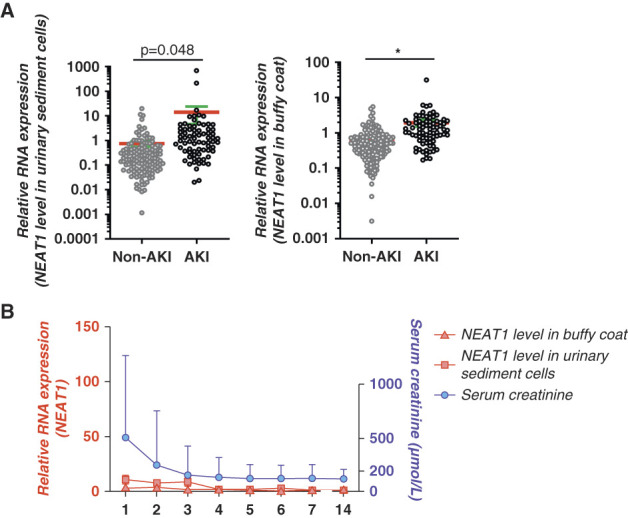
Results: In humans, hospitalized patients with AKI (N=66) demonstrated significantly higher lncRNA NEAT1 levels in urinary sediment cells and buffy coat versus control participants (N=152) from a primary care clinic; among six kidney transplant recipients, NEAT1 levels were the highest immediately after transplant surgery, followed by a prompt decline to normal levels in parallel with recovery of kidney function. In mice with AKI induced by sepsis (by LPS injection or cecal ligation and puncture) and renal ischemia-reperfusion, kidney tubular Neat1 was increased versus sham-operated mice. Knockdown of Neat1 in the kidney using short hairpin RNA preserved kidney function and suppressed overexpression of the AKI biomarker neutrophil gelatinase-associated lipocalin, leukocyte infiltration, and both intrarenal and systemic inflammatory cytokines IL-6, CCL-2, and IL-1β. In LPS-treated C1.1 cells, Neat1 was overexpressed by TLR4/NF-κB signaling and translocated from the cell nucleus into the cytoplasm where it promoted activation of nucleotide oligomerization domain-like receptor family protein 3 inflammasomes by binding with the scaffold protein receptor of activated protein C kinase 1. Silencing Neat1 ameliorated LPS-induced cell inflammation, whereas its overexpression upregulated IL-6 and CCL-2 expression even without LPS stimulation.

Conclusions: Our findings demonstrate a pathogenic role of NEAT1 induction in human and mice during AKI with alleviation of kidney injury in three experimental models of septic and aseptic AKI after knockdown of Neat1. LPS/TLR4-induced Neat1 overexpression in tubular epithelial cells increased the inflammatory response by binding with the scaffold protein, receptor of activated protein C kinase 1, to activate nucleotide oligomerization domain-like receptor family protein 3 inflammasomes.


Conflict of interest statement

Disclosure forms, as provided by each author, are available with the online version of the article at http://links.lww.com/JSN/E671.

Figures

None
Graphical abstract
Figure 1
Figure 1
Changes of NEAT1 levels in urinary sediment cells and buffy coat from patients with AKI. (A) Cross-sectional cohort among 66 hospitalized patients with AKI versus 152 control participants without AKI from a primary care clinic (data were presented in a logarithmic scale). (B) Longitudinal cohort (N=6) showing temporal changes in NEAT1 levels in relation to kidney function after kidney transplantation. *P < 0.001. NEAT1, nuclear-enriched abundant transcript 1.
Figure 2
Figure 2
Knockdown of Neat1 reduced AKI biomarkers and improved kidney function in sepsis-induced AKI mice. (A) Experimental design. Three days before LPS injection or CLP surgery, shNeat1 or empty plasmid (vehicle) was delivered into the kidney of C57BL/6J mice by tail vein injection, followed by ultrasound microbubble-mediated gene transfer. (B) Relative Neat1 expression in the kidney at 12 and 24 hours after LPS injection and 24 hours after CLP surgery (Ctl, n=3; LPS, n=6; Sham, n=3; CLP, n=6). (C) Representative ISH of Neat1 expression in the kidney and the corresponding quantitative analyses (Ctl, n≥2; LPS, n=4; Sham, n=3; CLP, n=6). Scale bar=100 μm. (D) Representative kidney morphology and the corresponding quantitative analyses (Ctl, n=3; LPS, n=6; Sham, n=3; CLP, n=6). Scale bar=100 μm. (E) Representative immunohistochemical staining of NGAL and the corresponding quantitative analyses (Ctl, n=3; LPS, n=6; Sham, n=3; CLP, n=6). Scale bar=100 μm. (F) Relative mRNA expression of NGAL in the kidney (Ctl, n=3; LPS, n=6; Sham, n=3; CLP, n=6). (G) Measurement of serum BUN and serum creatinine levels by ELISA (Ctl, n=3; LPS, n=6; Sham, n=3; CLP, n=6). All data are expressed as mean±SEM, *P < 0.001. CLP, cecal ligation and puncture; ctl, control; ISH, in situ hybridization; NGAL, neutrophil gelatinase–associated lipocalin; SCR, serum creatinine; shNeat1, shRNA plasmid targeting Neat1; shRNA, small hairpin RNA.
Figure 3
Figure 3
The renoprotective role of Neat1 knockdown in IR-induced AKI mice. (A) Experimental design. Three days before IR surgery, shRNA plasmid targeting Neat1 (shNeat1) or empty plasmid (vehicle) was delivered into the kidney of C57BL/6J mice by tail vein injection, followed by ultrasound microbubble-mediated gene transfer. (B) Relative Neat1 expression and representative ISH of Neat1 expression with the corresponding quantitative analyses in the kidney after IR surgery (Sham, n=3; IRI, n=6). (C) Representative morphology of the renal cortex and medulla (red arrowhead indicated injured areas) and the corresponding quantitative analyses (Sham, n=3; IRI, n=6). Scale bar=100 μm. (D) Representative immunohistochemical staining of NGAL with the corresponding quantitative analyses, and relative mRNA expression of NGAL in the kidney (Sham, n=3; IRI, n=6). Scale bar=100 μm. (E) Representative immunohistochemical staining of Ly6B.2 in the kidney interstitium (red arrowheads indicated positively stained cells) and the corresponding quantitative analyses (Sham, n=3; IRI, n=6). Scale bar=100 μm. (F) Measurement of serum BUN and serum creatinine levels by ELISA (Sham, n=3; IRI, n=6). (G) Relative mRNA expression of NLRP3, IL-1β, IL-6, and CCL-2 in the kidney (Sham, n=3; IRI, n=6). (H) Representative Western blot analyses of the expression levels of NLRP3, pro-caspase-1, and cleaved caspase-1 (p10) in the kidney after IR surgery, and the corresponding quantitative analyses (Sham, n=3; IRI, n=6). All data are expressed as mean±SEM, *P < 0.001. GAPDH, glyceraldehyde-3-phosphate dehydrogenase; IR, ischemia-reperfusion; IRI, ischemia-reperfusion injury; NLRP3, NOD-like receptor family protein 3; NOD, nucleotide oligomerization domain.
Figure 4
Figure 4
Knockdown of Neat1 attenuated kidney inflammation by suppressing NLRP3 inflammasome activation in sepsis-induced AKI mice. (A) Representative immunohistochemical staining of Ly6B.2 in the kidney interstitium (red arrowheads indicated positively stained cells) and the corresponding quantitative analyses (Ctl, n=3; LPS, n=6; Sham, n=3; CLP, n=6). Scale bar=100 μm. (B) Relative mRNA expression of NLRP3, IL-1β, IL-6, and CCL-2 in the kidney (Ctl, n=3; LPS, n=6; Sham, n=3; CLP, n=6). (C) Representative Western blot analyses of the expression levels of NLRP3, pro-caspase-1, and cleaved caspase-1 (p10) in the kidney at 12 hours after LPS injection and 24 hours after CLP surgery, and the corresponding quantitative analyses (Ctl, n=3; LPS, n=6; Sham, n=3; CLP, n=6). All data are expressed as mean±SEM, *P < 0.001.
Figure 5
Figure 5
Neat1 mediated inflammation in C1.1 mouse tubular cells. (A) Relative expression of Neat1, IL-6, and CCL-2 at different times after LPS stimulation by RT-qPCR (n=6). (B) Efficiency of Neat1 knockdown by Neat1 antisense locked nucleic acid GapmeR compared with negative control (Ctl) with or without LPS stimulation by RT-qPCR (n=6). Relative expression of IL-6 in cells transfected with Ctl or GapmeRs Neat1 under LPS stimulation as determined by (C) RT-qPCR, (D) Western blotting, and (E) ELISA (n=6). (F) Neat1 overexpression after transfection with pcDNA3.1 Neat1 plasmids by RT-qPCR (n≥6). Relative expression of IL-6 and CCL-2 in cells transfected with Ctl or Neat1 plasmids as determined by (F) RT-qPCR, (G) Western blotting, and (H) ELISA (n=6). All data are expressed as mean±SEM, *P < 0.001. pcDNA, plasmid cloning DNA; RT-qPCR, reverse transcription-quantitative PCR.
Figure 6
Figure 6
Neat1-mediated TLR4/NF-κB-induced NLRP3 inflammasome activation in C1.1 cells. (A) Schematic diagram of Neat1's promoter region on mouse chromosome 19. Primers were designed for the four putative p65/NF-κB binding sites (areas 1–4). (B) Enrichment of the p65/NF-κB binding fragment presented as percentage relative to input DNA. Anti-histone H3 antibody and normal rabbit IgG were used as positive and negative controls, respectively (n=3). (C) Relative expression of Neat1 under LPS stimulation with pretreatment of TLR4 inhibitor (CLI-095) or NF-κB inhibitors (BAY11-7085, JSH-23) by RT-qPCR (n≥5). (D) Representative Western blot analysis of expression levels of phospho-p65 and p65 in LPS-primed, nigericin-induced cells with Ctl or GapmeR Neat1 and the corresponding quantitative analyses (n=6). (E) Representative Western blot analysis of expression levels of pro-caspase-1 and cleaved caspase-1 (p10) in LPS-primed, nigericin-induced cells with Ctl or GapmeR Neat1 and the corresponding quantitative analyses (n≥5). (F) Western blotting showing the effect of Neat1 knockdown on ASC oligomerization in cell pellets from LPS-primed, nigericin-induced cells. Soluble cell lysates were used as input. (G) Representative immunofluorescence staining of endogenous ASC speck (red) and DAPI (blue) in LPS-primed, nigericin-induced cells with Ctl or GapmeR Neat1. Scale bar=50 μm. All data are expressed as mean±SEM, *P < 0.001. ASC, apoptosis-associated speck-like protein containing a CARD; CARD, caspase activation and recruitment domains; DAPI, 4',6-diamidino-2-phenylindole.
Figure 7
Figure 7
Neat1 interacts with Rack1 and regulates NLRP3 inflammasome. (A) Detection of Rack1 by Western blotting in RNA pull-down sample using a biotinylated Neat1 probe in LPS-stimulated C1.1 cells. The LacZ probe was used as negative control (n=3). (B) RIP using antibody against Rack1 or normal rabbit IgG (negative control) in LPS-stimulated C1.1 cell extracts followed by RT-qPCR with specific primers to Neat1 or U1 snRNA (negative control) (n=3). (C) Co-immunoprecipitation with anti-NLRP3 antibody on LPS-primed, nigericin-induced HK293T cells cotransfected with pcDNA3-Flag-NLRP3 and pcDNA3-Flag-ASC plasmids with control (Ctl) or Neat1 siRNA. Normal mouse IgG was used as negative control (n=3). (D) Representative Western blot analysis of the effect of Neat1 knockdown and Rack1 overexpression and (E) of the effect of Neat1 overexpression and Rack1 knockdown on expression of NLRP3, pro-caspase-1, cleaved caspase-1 (p10) and ASC in LPS-primed, nigericin-induced C1.1 cells and quantification (n≥5). All data are expressed as mean±SEM, *P < 0.001. pCMV, immediate early promoter of the human cytomegalovirus; Rack1, receptor of activated protein C kinase 1; RIP, RNA immunoprecipitation; siRNA, small interfering RNA; snRNA, small nuclear RNA.
Figure 8
Figure 8
Schematic diagram showing Neat1's role in NLRP3 inflammasome assembly triggered by LPS/nigericin.

References

    1. Vijayan A. Tackling AKI: prevention, timing of dialysis and follow-up. Nat Rev Nephrol. 2021;17(2):87–88. doi: 10.1038/s41581-020-00390-3 - DOI - PMC - PubMed
    1. Chan KW, Yu KY, Lee PW, Lai KN, Tang SC. Global REnal involvement of CORonavirus disease 2019 (RECORD): a systematic review and meta-analysis of incidence, risk factors, and clinical outcomes. Front Med (Lausanne). 2021;8:678200. doi: 10.3389/fmed.2021.678200 - DOI - PMC - PubMed
    1. Peerapornratana S, Manrique-Caballero CL, Gomez H, Kellum JA. Acute kidney injury from sepsis: current concepts, epidemiology, pathophysiology, prevention and treatment. Kidney Int. 2019;96(5):1083–1099. doi: 10.1016/j.kint.2019.05.026 - DOI - PMC - PubMed
    1. Arshad A, Ayaz A, Rehman S, Ukrani RD, Akbar I, Jamil B. Progression of acute kidney injury to chronic kidney disease in sepsis survivors: 1-year follow-up study. J Intensive Care Med. 2021;36(11):1366–1370. doi: 10.1177/0885066620956621 - DOI - PubMed
    1. Ronco C, Bellomo R, Kellum JA. Acute kidney injury. Lancet. 2019;394(10212):1949–1964. doi: 10.1016/S0140-6736(19)32563-2 - DOI - PubMed

MeSH terms

Substances

LinkOut - more resources