Review
. 2025 Jan;25(1-2):e2400022.
doi: 10.1002/pmic.202400022. Epub 2024 Aug 1.

Data acquisition approaches for single cell proteomics

Affiliations
Review

Data acquisition approaches for single cell proteomics

Gautam Ghosh et al. Proteomics. 2025 Jan.

Abstract

Single-cell proteomics (SCP) aims to characterize the proteome of individual cells, providing insights into complex biological systems. It reveals subtle differences in distinct cellular populations that bulk proteome analysis may overlook, which is essential for understanding disease mechanisms and developing targeted therapies. Mass spectrometry (MS) methods in SCP allow the identification and quantification of thousands of proteins from individual cells. Two major challenges in SCP are the limited material in single-cell samples necessitating highly sensitive analytical techniques and the efficient processing of samples, as each biological sample requires thousands of single cell measurements. This review discusses MS advancements to mitigate these challenges using data-dependent acquisition (DDA) and data-independent acquisition (DIA). Additionally, we examine the use of short liquid chromatography gradients and sample multiplexing methods that increase the sample throughput and scalability of SCP experiments. We believe these methods will pave the way for improving our understanding of cellular heterogeneity and its implications for systems biology.

Keywords: data dependent acquisition; data independent acquisition; mass spectrometry; multiplex; proteomics; single cell.


Conflict of interest statement

Brian C. Searle is a founder and shareholder in Proteome Software, which operates in the field of proteomics.

Figures

FIGURE 1
FIGURE 1
Comparison of MS spectra from different methods in single‐cell proteomics. (A) TMT‐DDA (Tandem Mass Tag with Data‐Dependent Acquisition) combines TMT labeling with data‐dependent acquisition, allowing for multiplexed quantification of proteins. In a TMT‐DDA MS2 spectrum, each peptide generates fragment ions. Alongside the fragment ions, specific reporter ions corresponding to each TMT channel are also observed. These reporter ions have unique masses that represent the different channels used for multiplexing. Meanwhile, the carrier ion helps increase sequence‐specific fragment ion intensity, which boosts the signal‐to‐noise ratio and enhances the detection of peptides during the analysis of the mass spectra. In this example, peptide signals have intensities of approximately 208 cells, where 200 come from the carrier channel combined with 8 single cells, as specified in the SCoPE method [62]. (B) LFQ‐DIA (Label‐Free Quantification with Data‐Independent Acquisition) relies on detecting and quantifying peptides between different single‐cell samples without using labels. Multiple peptide precursors can be fragmented simultaneously, typically resulting in highly complex MS2 spectra compared to DDA methods. (C) TMT‐DIA (Tandem Mass Tag with Data‐Independent Acquisition) multiplexes multiple samples within a single DIA experiment. One challenge of multiplexing both samples and peptides in the same measurement is that the same reporter ions in a TMT‐DIA spectrum can originate from multiple peptides, compromising their quantitative interpretation compared to TMT‐DDA. Without a carrier (as specified in Ctortecka et al. [77]), individual peptide signals would have a total intensity of 10 cells, whereas the MS2 signal can be the product of multiple peptides (e.g., 20 cells). (D) SIL‐DIA (Stable Isotope Labeling with Data‐Independent Acquisition) involves labeling proteins within cells with stable isotopes. In the MS2 spectra, N‐terminal b‐ions show distinct peaks corresponding to the different isotopic forms of the labeled peptides. These peaks are separated by the mass difference between the heavy isotopes. Meanwhile, C‐terminal y‐type ions may or may not be labeled depending on whether they contain a lysine, which results in y‐type ion peaks that are the combined intensities of all three isotopic variants.
FIGURE 2
FIGURE 2
A comparison of traditional stable isotope labeling methods (like SILAC) and amine‐reactive tagging methods (like mTRAQ) for quantitative proteomics. (A) Stable isotope labeling first involves labeling cells in culture with stable isotopes of amino acids in three distinct forms: light (naturally occurring isotopic composition), medium (partially labeled), and heavy (fully labeled). Using this modality, peptides are labeled on the c‐terminus, which produces isomeric y‐type ions that indicate the quantity of each sample. (B) In contrast, mTRAQ utilizes nonisobaric tags to label peptides individually after cells are cultured, lysed, and digested. The labeled peptides are then combined for MS analysis. Here, peptides are labeled at free amines, shifting the masses of b‐type ions, which are typically more unstable and less useful for quantification. While some peptides that terminate in lysines will also have y‐type ion labels, typically quantification is performed at the precursor level, rather than the fragment level.
FIGURE 3
FIGURE 3
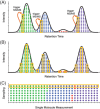
A comparison of DDA, DIA, and single‐molecule methods in SCP. (A) DDA attempts to make a single measurement of each precursor ion once the precursor intensity achieves a specific threshold. While this approach increases the number of detected peptides by avoiding measuring any given peptide multiple times, it limits the total amount of actual ions used to measure that peptide (orange boxes). Also, there is a stochastic chance that low abundance peaks never achieve the threshold and may be missed due to dynamic exclusion, which can limit sensitivity with low‐input samples. (B) DIA continuously samples a predefined mass range, which means that each peptide is sampled near its apex, leading to a higher likelihood of detecting low‐abundance peptides. (C) Single‐molecule measurements introduce the possibility of missing low‐abundance proteins because many highly abundant molecules must be sampled before the chance of observing low‐abundant molecules becomes a certainty.

Similar articles

Cited by

References

    1. Regev, A. , Teichmann, S. A. , Lander, E. S. , Amit, I. , Benoist, C. , Birney, E. , Bodenmiller, B. , Campbell, P. , Carninci, P. , Clatworthy, M. , Clevers, H. , Deplancke, B. , Dunham, I. , Eberwine, J. , Eils, R. , Enard, W. , Farmer, A. , Fugger, L. , Göttgens, B. , … The Human Cell Atlas . (2017). The human cell atlas. eLife, 6. 10.7554/eLife.27041 - DOI - PMC - PubMed
    1. Shlush, L. I. , Mitchell, A. , Heisler, L. , Abelson, S. , Ng, S. W. K. , Trotman‐Grant, A. , Medeiros, J. J. F. , Rao‐Bhatia, A. , Jaciw‐Zurakowsky, I. , Marke, R. , Mcleod, J. L. , Doedens, M. , Bader, G. , Voisin, V. , Xu, C. , Mcpherson, J. D. , Hudson, T. J. , Wang, J. C. Y. , Minden, M. D. , … Dick, J. E. (2017). Tracing the origins of relapse in acute myeloid leukaemia to stem cells. Nature, 547(7661), 104–108. - PubMed
    1. Nam, A. S. , Kim, K. T. , Chaligne, R. , Izzo, F. , Ang, C. , Taylor, J. , Myers, R. M. , Abu‐Zeinah, G. , Brand, R. , Omans, N. D. , Alonso, A. , Sheridan, C. , Mariani, M. , Dai, X. , Harrington, E. , Pastore, A. , Cubillos‐Ruiz, J. R. , Tam, W. , Hoffman, R. , … Landau, D. A. (2019). Somatic mutations and cell identity linked by Genotyping of Transcriptomes. Nature, 571(7765), 355–360. - PMC - PubMed
    1. Kreso, A. , & Dick, J. E. (2014). Evolution of the cancer stem cell model. Cell Stem Cell, 14(3), 275–291. - PubMed
    1. Ramsköld, D. , Luo, S. , Wang, Y.‐C. , Li, R. , Deng, Q. , Faridani, O. R. , Daniels, G. A. , Khrebtukova, I. , Loring, J. F. , Laurent, L. C. , Schroth, G. P. , & Sandberg, R. (2012). Full‐length mRNA‐Seq from single‐cell levels of RNA and individual circulating tumor cells. Nature Biotechnology, 30(8), 777–782. - PMC - PubMed

LinkOut - more resources